Ashley T. Gault, DO1, Kashif Tufail, MD2, Krishna Kothamasu, DO1, Huzaif Taufiq, MD3 1Jefferson Torresdale Hospital, Philadelphia, PA; 2Northeast Gastroenterology Associates, Philadelphia, PA; 3Jefferson Torresdale Hospital, Willow Grove, PA Introduction: This case involves rare, rapidly progressing necrotizing pancreatitis with complications including splenic vein thrombosis, intraperitoneal hemorrhage, and abdominal compartment syndrome. While each of these issues can happen on their own in acute pancreatitis, having them all occur one after another, and so quickly, led to severe instability and multi-organ failure, making this a particularly dangerous and unusual case.
Case Description/
Methods: A 60-year-old female with a history of hypertension, hyperlipidemia, type 2 diabetes, and cholecystectomy (2010) presented to the emergency room with 3 days of sharp, localized epigastric pain, bilious vomiting, and watery diarrhea. She denies fever, chest pain, flank pain, urinary symptoms, alcohol use, or recent surgeries.
Initially hemodynamically stable, she had epigastric tenderness and labs showed elevated lipase, transaminitis, and leukocytosis. CT showed peripancreatic inflammation and possible splenic vein thrombosis. Doppler ultrasound confirmed splenic vein occlusion. She was managed with fluids, pain control, and started on a heparin infusion. She was admitted for pancreatitis of unclear etiology, complicated by splenic vein thrombosis. Her diet was slowly advanced with occasional epigastric pain, until a rapid response occurred one week later.
She developed encephalopathy, hypotension, tachycardia, and a hemoglobin dropped to 5.6. Heparin was reversed, massive transfusion protocol initiated, and she was intubated and transferred to the ICU. Once stable, CTA showed no active bleed, but abdominal distension and firmness prompted emergent surgery. She underwent decompressive laparotomy with evacuation of 2L of blood and splenic artery embolization.
Nine days post-op, she was extubated, tolerated diet advancement, and resumed normal bowel function. After a month-long hospital stay, she was discharged home in stable condition. Discussion: This case highlights the critical importance of early recognition of complications in severe pancreatitis. Careful, ongoing assessment, including timely imaging in response to changes in pain or clinical status, is key to identifying abdominal necrosis. Acting quickly can help prevent serious complications like hemorrhage and abdominal compartment syndrome. Overall, this case underscores the need for close clinical attention and a well-coordinated, multidisciplinary approach when managing complex pancreatitis cases.
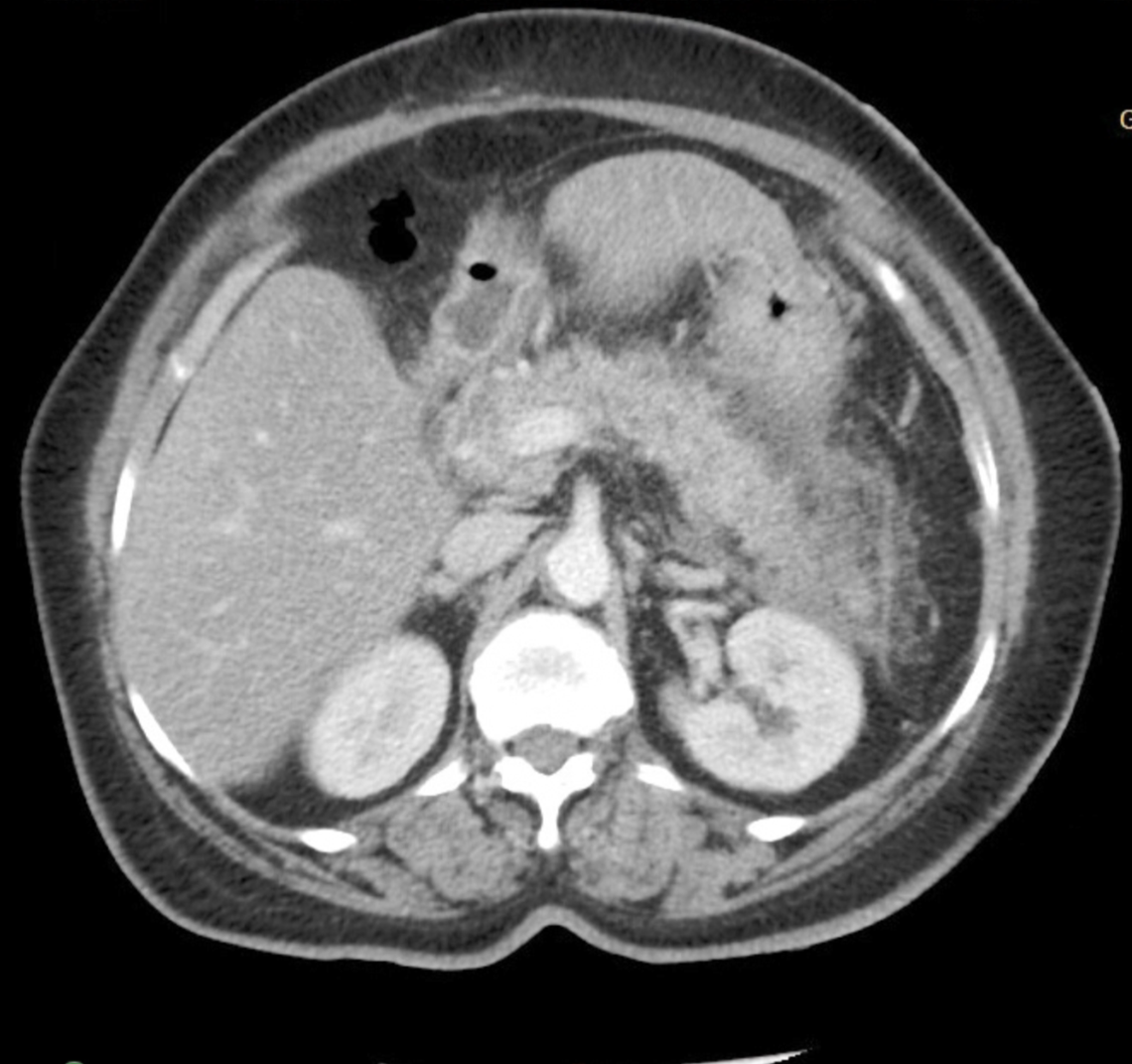
Figure: CT abdomen/pelvis (8/27/2024) Acute pancreatitis: Peripancreatic inflammatory stranding with mild adjacent fluid, but no focal rim- enhancing collection. Suspected thrombosis of portions of the splenic vein.
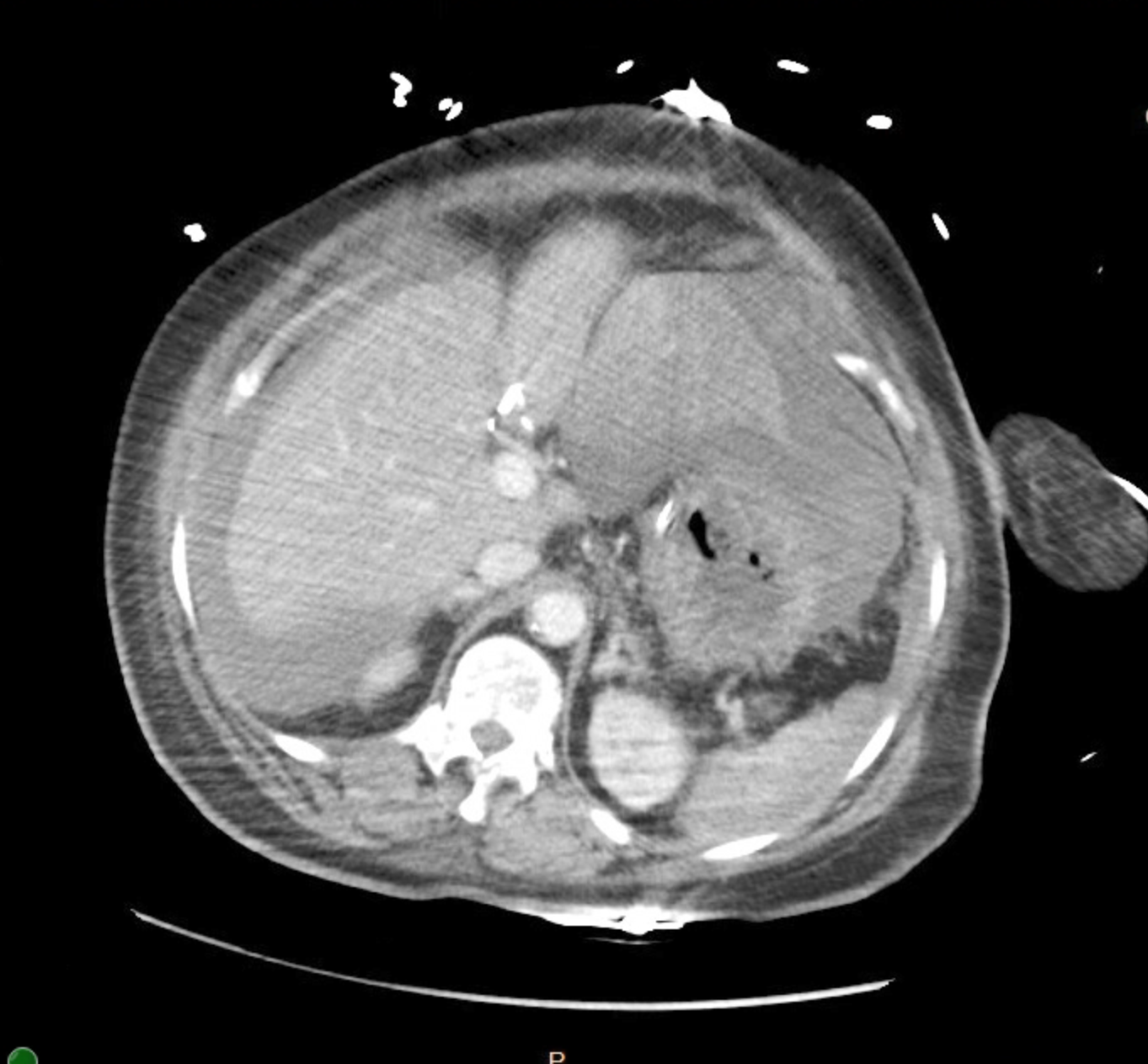
Figure: CTA abdomen/pelvis (9/3/2024) Hemoperitoneum without definite active arterial bleeding: Hyperdense fluid layering anterior to the left hepatic lobe and along the greater curvature of the stomach, possibly pooling in the lesser sac or anterior peritoneal space, suggesting subacute hemorrhage. Ill- defined areas of hyperdensity in the omentum suggesting small areas of omental hemorrhage.
Disclosures: Ashley Gault indicated no relevant financial relationships. Kashif Tufail indicated no relevant financial relationships. Krishna Kothamasu indicated no relevant financial relationships. Huzaif Taufiq indicated no relevant financial relationships.
Ashley T. Gault, DO1, Kashif Tufail, MD2, Krishna Kothamasu, DO1, Huzaif Taufiq, MD3. P4464 - Bleeding Out Inside: When Pancreatitis Turns Deadly, ACG 2025 Annual Scientific Meeting Abstracts. Phoenix, AZ: American College of Gastroenterology.