Matthew Posen, DO, Krishna Shah, MD, Ahmad Mardini, MBBS, Ernst Joachim, MD, Samee Farooqi, DO, Tarek Almouradi, MD Advocate Christ Medical Center, Oak Lawn, IL Introduction: Solitary fibrous tumors (SFT) are a rare mesenchymal neoplasm most commonly seen in the pleura that can occur throughout the body. These tumors are often found incidentally and have only been reported to be in the pancreas less than fifty times. Here, we present an example of this very rare tumor presentation.
Case Description/
Methods: An 83-year-old woman with history of dementia presented to the hospital with complaint of shortness of breath for two weeks of duration. Computerized tomography (CT) angiography chest was ordered. No pulmonary embolism was visualized; however, the imaging did incidentally reveal suggestion of a mass in the body of the pancreas measuring 2.0x2.3cm with punctate calcifications. Magnetic resonance imaging (MRI) abdomen demonstrated a hyperenhancing heterogeneous mass of the pancreatic body measuring 2.7cm. Endoscopic ultrasound with biopsy was completed with histology demonstrating fibrotic tissue with spindle cell proliferation and immunostaining positive for CD34 and STAT6. Final diagnosis of solitary fibrous tumor of the pancreas was made with plan to follow up outpatient for observation without plan for resection. Discussion: The first reported case of pancreatic SFT was as recent as 1999 with just over 40 cases reported since. A 2023 literature review of forty-four reported cases of pancreatic SFT’s analyzed typical imaging, histology, and presentation. Imaging for these lesions typically describes them as hypervascular, multinodular, solid masses. Histology shows patternless distributions of ovoid to spindle shaped cells. Testing for STAT6 and CD34 were typically positive with additionally high rates for bcl-2. Most cases were benign with 10-15% of cases acting aggressively with local recurrence or distant metastasis. Another study analyzed 110 cases of SFTs of non-specific locations to create a 3-tiered risk assessment model for metastasis. Poor prognostic markers included age greater than 55, tumor size greater than 5cm, and mitotic figures greater than 4 power fields. Low-risk patients had no metastasis or death secondary to SFTs on median follow up of 50 months. The five-year disease-specific mortality rate in the moderate and high-risk groups was 7% and 40% respectively. Thus, it is not unreasonable for low-risk patients with comorbidities to forgo surgical resection and instead opt for close observation and follow-up. Much work remains to be done to elucidate management and potential risk factors of SFTs.
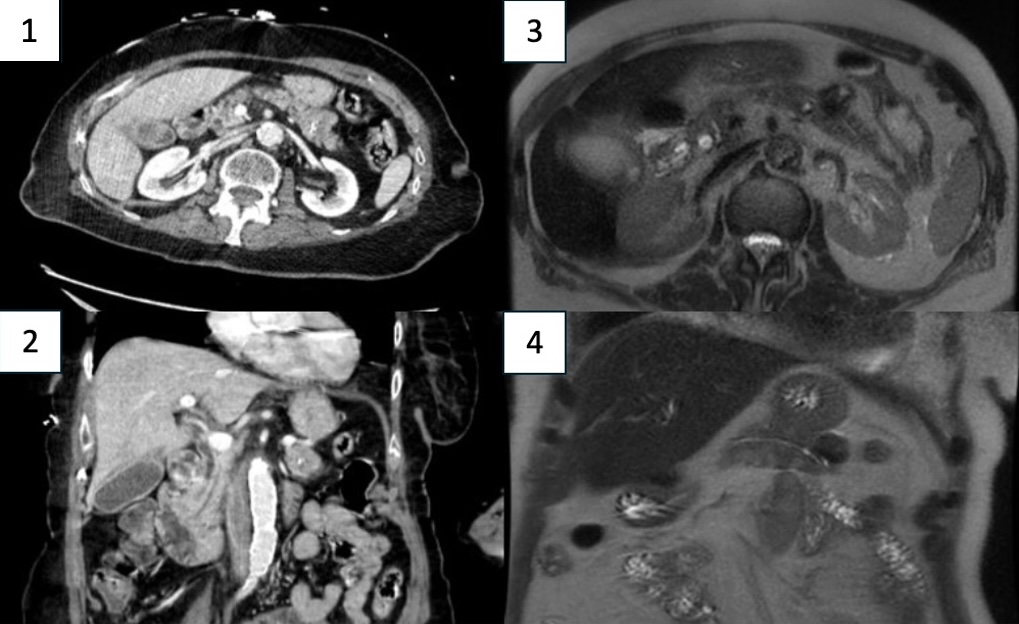
Figure: Image 1: CT Abdomen Pelvis demonstrating a 2.4cm x 2.1cm pancreatic mass with internal calcification in the transverse view. Image 2: CT Abdomen Pelvis demonstrating a 2.4cm x 2.1cm pancreatic mass with internal calcification in the coronal view. Image 3: MRI Abdomen redemonstrating the pancreatic mass in the transverse view. Image 4: MRI Abdomen redemonstrating the pancreatic mass in the coronal view.
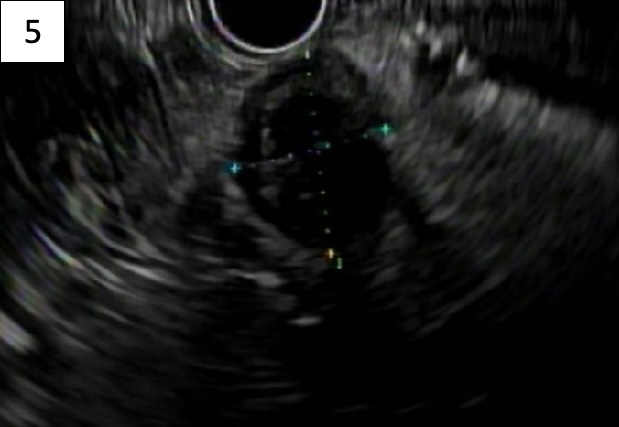
Figure: Image 5: Endoscopic ultrasound showing a well-demarcated, heterogenous, mainly hypoechoic pancreatic body mass measuring approximately 2cm x 2cm.
Disclosures: Matthew Posen indicated no relevant financial relationships. Krishna Shah indicated no relevant financial relationships. Ahmad Mardini indicated no relevant financial relationships. Ernst Joachim indicated no relevant financial relationships. Samee Farooqi indicated no relevant financial relationships. Tarek Almouradi indicated no relevant financial relationships.
Matthew Posen, DO, Krishna Shah, MD, Ahmad Mardini, MBBS, Ernst Joachim, MD, Samee Farooqi, DO, Tarek Almouradi, MD. P4454 - Solitary Fibrous Tumor of the Pancreas: An Exceedingly Rare Tumor Presentation, ACG 2025 Annual Scientific Meeting Abstracts. Phoenix, AZ: American College of Gastroenterology.