Essam Rashad, MBBCh1, Vinay Katukuri, MD2 1Parkview Medical Center, Fort Wayne, IN; 2Advanced Gastroenterology of Central Florida, Orlando, FL Introduction: Pancreatic cystic lymphangiomas account for less than 1% of all lymphangiomas and less than 0.2% of pancreatic cysts. First described by Koch in 1913, fewer than 100 cases have been reported worldwide. Diagnosis has traditionally required surgical resection and histopathological examination, though endoscopic ultrasound with fine needle aspiration (EUS-FNA) has emerged as a valuable diagnostic tool. We present a case of pancreatic cystic lymphangioma diagnosed and managed by EUS and FNA, avoiding the need for surgery.
Case Description/
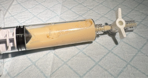
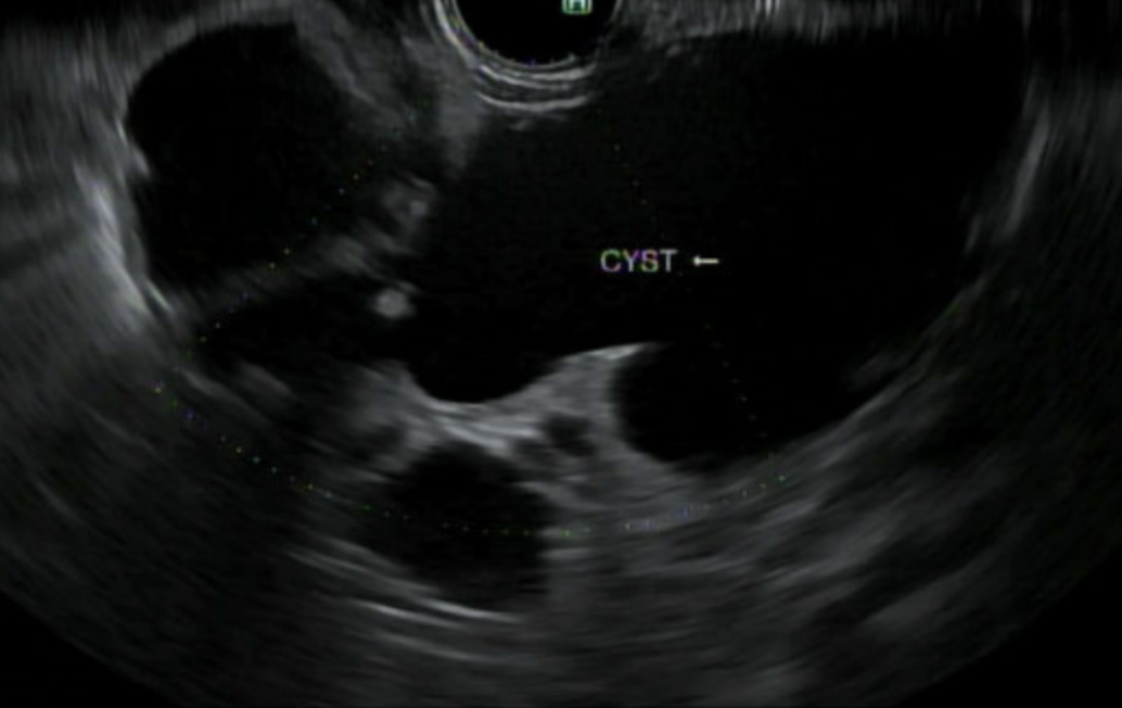
Methods: A 54-year-old male with ulcerative colitis underwent routine MR enterography, revealing an incidental 6.8 cm septated cystic lesion arising from the pancreatic head without main pancreatic duct dilatation or enhancement. EUS demonstrated anechoic lesions with two large compartments surrounded by small cystic areas. Fine needle aspiration yielded 100 mL of white, milky fluid. Laboratory analysis revealed extremely elevated triglycerides (6000 mg/dL), CEA 1.2 ng/mL, and amylase 153 U/L. Cytology was negative for malignant cells. The combination of chylous fluid appearance and markedly elevated triglycerides confirmed the diagnosis of pancreatic lymphangioma. Discussion: Lymphangiomas are congenital benign hamartomas arising from the lymphatic system. Pancreatic cystic lymphangiomas account for less than 1% of all lymphangiomas and < 5% of all pancreatic neoplasms. These lesions occur more frequently in females and are typically located in the pancreatic body or tail. Symptoms most commonly include abdominal pain and/or are a result of mass effect on nearby organs, though many cases are discovered incidentally during imaging for unrelated conditions. Cross-sectional imaging findings may resemble IPMN, mucinous or serous cystadenomas, and pseudocysts, necessitating sampling. Histology shows epithelial cells positive for Factor VIII-R antigen and CD31. Management options include cystectomy, distal pancreatectomy, and pancreaticoduodenectomy, depending on the size and location of the lesion and associated symptoms; however, EUS with aspiration and drainage is emerging as a potentially safe alternative approach. Further research is necessary to optimize management given the increasing utilization of EUS and advanced endoscopic techniques.
Figure: Multiple cysts in the body of the pancreas with largest 47 x 25 mm in diameter.
Figure: 10 ml syringe with triglyceride-rich white fluid characteristic of lymphangioma.
Disclosures: Essam Rashad indicated no relevant financial relationships. Vinay Katukuri indicated no relevant financial relationships.
Essam Rashad, MBBCh1, Vinay Katukuri, MD2. P4410 - Pancreatic Cystic Lymphangioma: A Rare Case Report, ACG 2025 Annual Scientific Meeting Abstracts. Phoenix, AZ: American College of Gastroenterology.