Texas Tech University Health Science Center El Paso El Paso, TX
Mutaz Kalas, MD1, M. Ammar Kalas, MD2, Shaked Laks, MD2, Akanksha Togra, MD3, Ioannis Konstantinidis, MD4, Sherif E. Elhanafi, MD2 1Texas Tech University Health Science Center El Paso, El Paso, TX; 2Texas Tech University Health Sciences Center, El Paso, TX; 3Texas Tech University Health Sciences Center, El Paso, El Paso, TX; 4Texas Oncology, El Paso, TX Introduction: Necrotizing pancreatitis (NP) is a severe and potentially life-threatening condition that is commonly due to benign underlying etiologies. PDAC is rarely identified in the setting of acute necrotizing pancreatitis with walled-off pancreatic collection (WOPN). This is a rare entity that may delay the diagnosis of cancer and is only reported in a few cases in the literature. Our case describes a 57-year-old male with acute NP and WOPN requiring cystgastrostomy with multiple necrosectomies, and was subsequently diagnosed with PDAC.
Case Description/
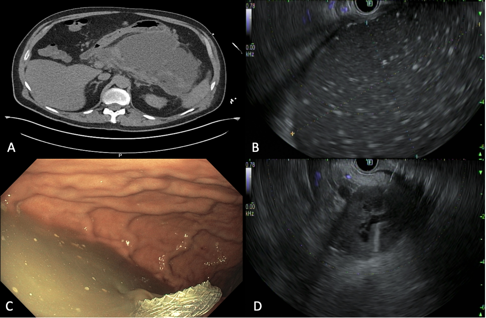
Methods: A 57-year-old male with a past medical history of type 2 diabetes mellitus, hypertension, and a history of alcohol use disorder presented to the ED with severe abdominal pain radiating to the back and associated with nausea and two episodes of non-bloody emesis. On exam, he was tachycardic, with epigastric tenderness and fullness on palpation. Computed tomography (CT) of the abdomen showed multiple fluid collections in the head and body of the pancreas, with the largest measuring 18 x 11 x 12 cm, displacing the stomach anteriorly. Subsequently, the patient underwent upper endoscopic ultrasonography (EUS) with cystgastrostomy and AXIOS stent placement. The patient underwent multiple endoscopic necrosectomies over the span of 2 months prior to removal of the AXIOS stent. Follow-up imaging showed improvement and resolution of WOPN with residual small collections in the head and body of the pancreas. Few months later, the patient reported recurrent abdominal pain which led to obtaining cross-sectional imaging, which revealed an ill-defined mass in the head of the pancreas. Decision was made to proceed with EUS for better characterization of the lesion, EUS showed a 4.3cm by 3.9cm hypoechoic, heterogenous mass in the head and uncinate process of the pancreas with upstream pancreatic duct dilation. Fine needle biopsy was performed, which revealed PDAC. CA19-9 was 371, and the patient is currently undergoing neoadjuvant chemotherapy. Discussion: The presentation of PDAC as acute NP with large WOPN is rare and can be challenging to diagnose, especially in the setting of WOPN, which may obscure the visualization of malignancy on cross-sectional imaging and endoscopic ultrasonography. This case highlights the importance of considering malignancy as one of the potential causes of acute NP with WOPN, particularly in patients with symptom recurrence after adequate drainage and treatment of the WOPN.
Figure: A) CT abdomen: large fluid collection measuring up to 18 x 11 x 12 cm B) EUS: large pseudocyst with areas of necrosis, C) Fluid draining following cystgastrostomy with AXIOS stent placement, D) EUS: 43 mm by 39 mm hypoechoic, heterogenous mass was identified in the pancreatic head and uncinate process of the pancreas with upstream pancreatic duct dilation
Disclosures: Mutaz Kalas indicated no relevant financial relationships. M. Ammar Kalas indicated no relevant financial relationships. Shaked Laks indicated no relevant financial relationships. Akanksha Togra indicated no relevant financial relationships. Ioannis Konstantinidis indicated no relevant financial relationships. Sherif Elhanafi indicated no relevant financial relationships.
Mutaz Kalas, MD1, M. Ammar Kalas, MD2, Shaked Laks, MD2, Akanksha Togra, MD3, Ioannis Konstantinidis, MD4, Sherif E. Elhanafi, MD2. P4378 - Necrotizing Pancreatitis With Walled-Off Necrosis: Beneath the Chaos, Malignancy May Be Masked, ACG 2025 Annual Scientific Meeting Abstracts. Phoenix, AZ: American College of Gastroenterology.