Murad H.. Ali, MD, Jane Lee, MD, David Friedel, MD NYU Langone Health, Mineola, NY Introduction: A 73-year-old male with a history of hypertension, chronic renal insufficiency, benign prostatic hyperplasia, gout, resected melanoma and psoriasis presented for evaluation of a distal small bowel lesion and iron deficiency anemia. Upper endoscopy and colonoscopy were unremarkable. Capsule endoscopy revealed a polypoid lesion in the mid- ileum, which was confirmed as an 18 mm polyp by CT enterography. A laparoscopic resection was initially considered.
Leiomyomas are common throughout the gastrointestinal (GI) tract but are rarely ulcerated and typically not clinically significant. When ulcerated, these lesions can become symptomatic, leading to complications such as chronic bleeding and iron deficiency anemia. In this patient, the leiomyoma caused significant iron deficiency anemia, prompting the need for evaluation and treatment via deep enteroscopy.
Case Description/
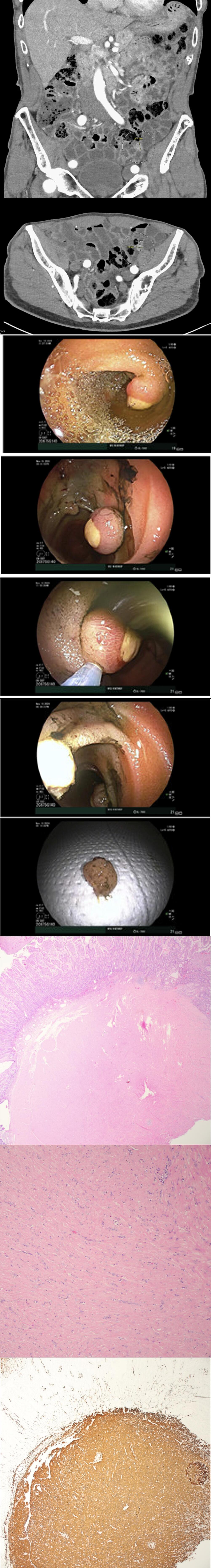
Methods: CT enterography revealed an 18 mm polyp in the mid ileum. Double-balloon enteroscopy (DBE) was performed to assess the lesion. A semi-pedunculated, ulcerated 18 mm polyp was identified in the mid ileum, with evidence of friability and slight bleeding. The lesion was resected using a hot snare, and the area was tattooed with 3 mL of Spot (carbon black) for future surgical localization. Epinephrine was injected for hemostasis. The remainder of the exam was normal. The procedure was well tolerated and the patient discharged.
The patient had significant iron deficiency anemia pre-procedure, as evidenced by low hemoglobin (11.3 g/dL), low iron (24 µg/dL), low ferritin (29 ng/mL), and a low iron saturation (9%). Post-procedure, the patient's lab results showed a marked improvement in hemoglobin (13.9 g/dL), iron (97 µg/dL), ferritin (118 ng/mL), and iron saturation (38%), indicating successful resolution of the anemia after the resection of the leiomyoma. As a result, surgical small bowel resection was avoided for patient and he was extremely grateful for this.
Discussion: Anterograde deep enteroscopy was effective in diagnosing and managing the distal small bowel leiomyoma in this patient with iron deficiency anemia. This case underscores the role of DBE in identifying and resecting rare small bowel lesions and highlights its utility in facilitating surgical management. The improvement in hemoglobin and iron levels post-procedure further emphasizes the importance of addressing underlying small bowel lesions in patients with unexplained iron deficiency anemia.
Figure: Images 1 & 2: CT enterography findings of ileal lesion Images 3/4/5/6/7: DBE findings of semi-pedunculated, ulcerated 18 mm polyp was identified in the mid ileum, with polypectomy and specimen retrieval Images 8/9: Ileum with leiomyoma and focal pyloric gland metaplasia. Image 10: The spindle cells are diffusely positive for desmin and negative for S100 and CD117, supporting the diagnosis of Leiomyoma.
Disclosures: Murad Ali indicated no relevant financial relationships. Jane Lee indicated no relevant financial relationships. David Friedel indicated no relevant financial relationships.
Murad H.. Ali, MD, Jane Lee, MD, David Friedel, MD. P1976 - Small Bowel, Big Impact: Deep Enteroscopy With Ileal Polypectomy of Ulcerated Pedunculated Leiomyoma Resolving Iron Deficiency Anemia, ACG 2025 Annual Scientific Meeting Abstracts. Phoenix, AZ: American College of Gastroenterology.