Alexander Siegel, MD1, Kyaw Min Tun, DO2, Wuttiporn Manatsathit, MD2, Jennifer Gamache, MD3 1CHI Health Creighton University Medical Center, Elkhorn, NE; 2Creighton University Medical Center, Omaha, NE; 3Creighton University, Omaha, NE Introduction: Strongyloidiasis, is a parasitic infection endemic to warm, and moist climates, that is caused by the intestinal nematode Strongyloides stercoralis. In the United States, most cases are seen in immunocompromised patient’s residing in the Northeast and East South Central regions and travelers from endemic regions. While often underrecognized, Strongyloides infection should remain a key consideration in patients presenting with atypical gastrointestinal (GI) symptoms, especially with a relevant exposure history.
Case Description/
Methods: A 45-year-old Hispanic immigrant male with no pertinent past medical history presented to gastroenterology clinic with a two-month history of hematochezia associated with epigastric pain. Rectal exam notable for hemorrhoids with normal hemoglobin of 14.6 g/dL. Heliobacter pylori breath test was positive, and patient treated with quadruple therapy. Four months later, outpatient upper endoscopy demonstrated non-bleeding duodenal erosions with erythematous gastric mucosa. Biopsies revealed normal gastric mucosa without H. pylori, but no duodenal biopsy taken. Colonoscopy notable for hemorrhoids with random colon biopsies showing active colitis with eosinophilic infiltrates, granulomas and focal cryptitis. The patient did not follow up afterwards due to lack of insurance. He then presented six months later to the hospital with right upper quadrant abdominal pain and nausea. Labs notable for leukocytosis of 22.1 K/uL with eosinophilia, alanine aminotransferase 237 u/L, aspartate aminotransferase 74 u/L, and hemoglobin 19.9 g/dL. Colonoscopy was grossly normal with similar histologic findings as prior. Upper endoscopy revealed evidence of duodenitis with biopsy showing villous atrophy, eosinophils and parasitic structures within the crypts consistent with Strongyloides stercoralis. The patient was treated with Ivermectin for two days. However, due to the patient’s lack of insurance and a functional phone number, a follow up appointment could not be arranged. Discussion: This case highlights the diagnostic complexity of chronic GI symptoms without a definitive diagnosis. It remains important to maintain a wide differential in patients with unexplained GI symptoms, especially when risk factors for parasitic infections are present.Strongyloidiasis often persists for years due to the parasite’s ability to auto-infect their host, allowing continuous reinfection without further exposure. Early tissue diagnosis is critical to avoid misdiagnosis and ensure correct treatment.
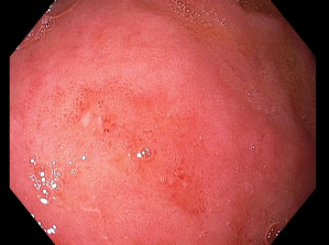
Figure: Endoscopic image of duodenal bulb with non-bleeding mucosal erosions
Figure: H&E slides of duodenal biopsy demonstrating duodenitis with villous atrophy and increased intramucosal eosinophils. At higher magnification (right), a structure can be seen in one of the crypts that has features of a nematode such as Strongyloides stercoalis.
Disclosures: Alexander Siegel indicated no relevant financial relationships. Kyaw Min Tun indicated no relevant financial relationships. Wuttiporn Manatsathit indicated no relevant financial relationships. Jennifer Gamache indicated no relevant financial relationships.
Alexander Siegel, MD1, Kyaw Min Tun, DO2, Wuttiporn Manatsathit, MD2, Jennifer Gamache, MD3. P1962 - Parasitic Persistence: The Diagnostic Challenge of Strongyloidiasis in a Non-Endemic Setting, ACG 2025 Annual Scientific Meeting Abstracts. Phoenix, AZ: American College of Gastroenterology.