Allison Malick, MD1, Shannon Todd, PA-C, MHS, MPAS2, Ahmad Alomari, MD3, Syed-Mohammed Jafri, MD3 1Henry Ford Hospital, Detroit, MI; 2Henry Ford Hospital Transplant Institute, Detroit, MI; 3Henry Ford Health, Detroit, MI Introduction: Pelvic Congestion Syndrome (PCS) is a chronic pain disorder often caused by venous insufficiency within the pelvic veins, most commonly attributed to hormonal and structural factors. PCS secondary to portosystemic shunting is a rare and underrecognized etiology. We present a case of PCS in a patient with non-alcoholic steatohepatitis (NASH)-related cirrhosis, caused by retrograde pelvic venous flow through an ovarian portosystemic shunt, without other manifestations of portal hypertension.
Case Description/
Methods: A 52-year-old female with NASH-related cirrhosis complicated by encephalopathy presents with several months of progressive pelvic pain. She was previously listed for liver transplant, but this is deferred after recompensation. She reports persistent, cramping pelvic discomfort worsened by standing, sitting, or twisting, as well as pelvic pressure and pain with toileting and intercourse. Physical examination reveals mild lower abdominal tenderness. Laboratory values show total bilirubin 1.1 mg/dL, ALP 144 U/L, ALT 22 U/L, AST 34 U/L. Contrast-enhanced CT of the abdomen and pelvis reveals dilated ovarian and uterine veins, including marked dilation and tortuosity of the right ovarian vein near the inferior vena cava (IVC), as well as enlargement of the right inferior mesenteric and splenic veins. The IVC is dilated (4.7 cm). A portosystemic shunt venogram demonstrates retrograde flow into the pelvic ovarian veins, with a large ovarian–to–internal iliac vein shunt, consistent with PCS. Notably, the patient lacks other signs of portosystemic shunting, including esophageal varices, splenomegaly, ascites, or rectal varices. Interventional Radiology performs coil embolization of two branches of the right ovarian vein, resulting in resolution of retrograde pelvic flow. Follow-up imaging shows decreased caliber of the parauterine vessels, and the patient reports approximately 75% improvement in pelvic pain. Discussion: This case highlights an unusual presentation of abdominal pain in a patient with cirrhosis and an uncommon vascular cause of PCS due to portosystemic shunting. The absence of other manifestations of portal hypertension makes the diagnosis particularly challenging. This case underscores the importance of considering central venous hypertension as a mechanism of pelvic pain in cirrhotic patients. It also illustrates the role of multidisciplinary evaluation, including imaging and endovascular intervention, in diagnosing and treating atypical manifestations of portal hypertension.
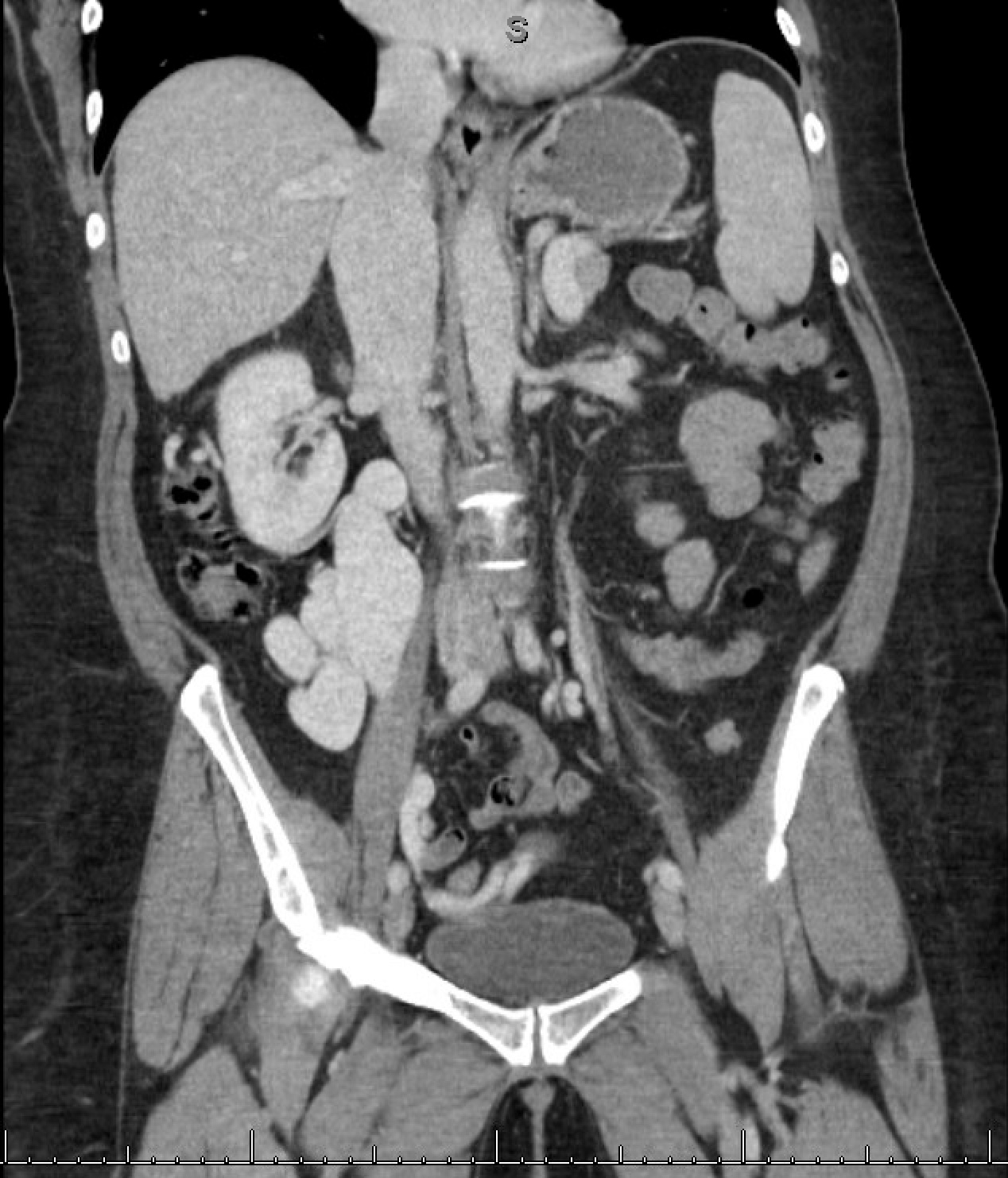
Figure: Contrast-enhanced CT of the abdomen and pelvis reveals dilated ovarian and uterine veins, including marked dilation and tortuosity of the right ovarian vein near the inferior vena cava (IVC), as well as enlargement of the right inferior mesenteric and splenic veins.
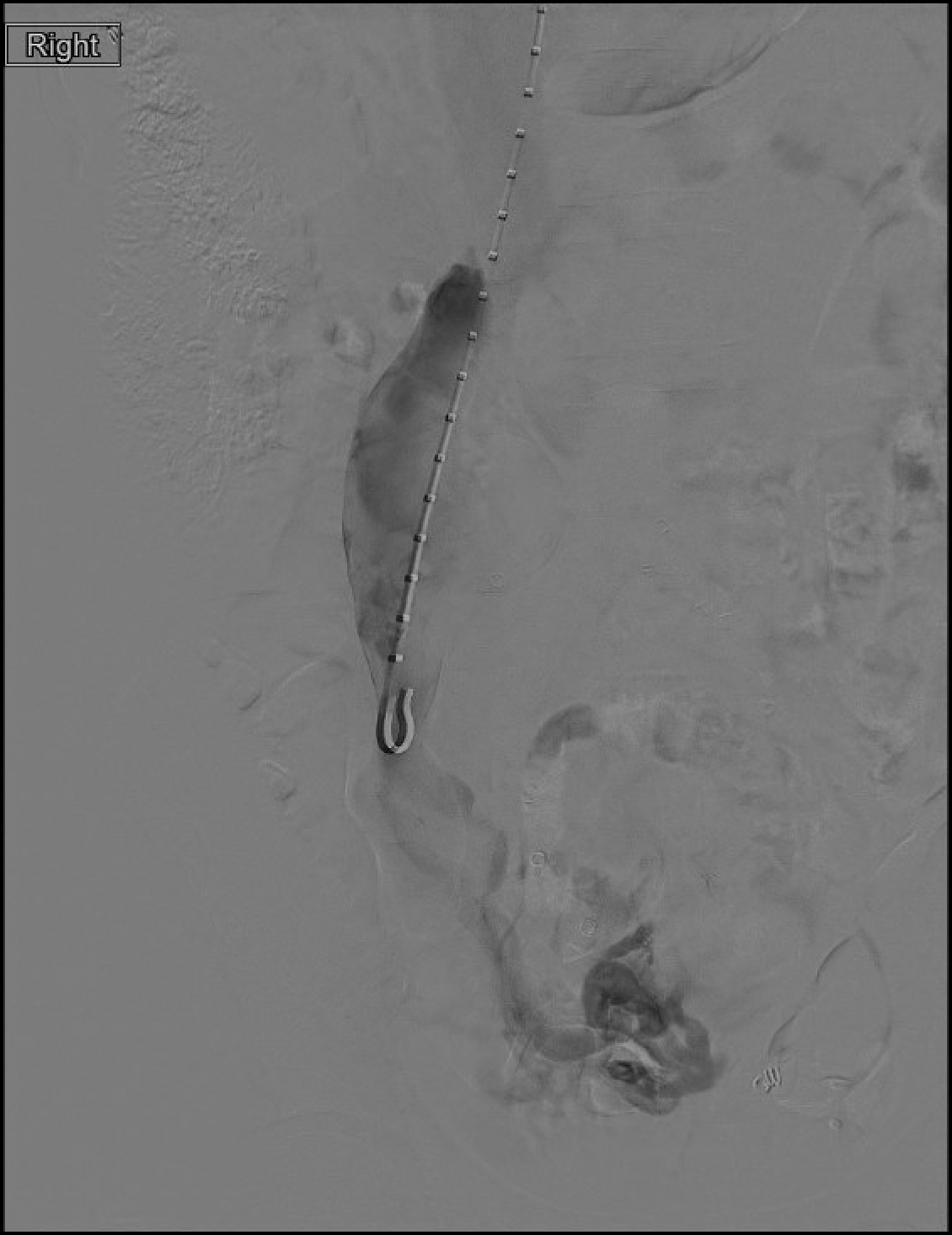
Figure: A portosystemic shunt venogram demonstrates retrograde flow into the pelvic ovarian veins, with a large ovarian–to–internal iliac vein shunt.
Allison Malick, MD1, Shannon Todd, PA-C, MHS, MPAS2, Ahmad Alomari, MD3, Syed-Mohammed Jafri, MD3. P1836 - An Unusual Presentation of Pelvic Congestion Syndrome Due to Spontaneous Portosystemic Shunting in a Cirrhotic Patient, ACG 2025 Annual Scientific Meeting Abstracts. Phoenix, AZ: American College of Gastroenterology.

 photo")