Allegheny Health Network Medicine Institute Pittsburgh, PA
Hany Habib, MD1, Khaldoun Arabi, MD2, Anas Alsolaiman, MD3, Samir Haffar, MD4 1Allegheny Health Network Medicine Institute, Pittsburgh, PA; 2Damascus University, Regensburg, Bayern, Germany; 3Abbasieen Hospital, Damascus, Dimashq, Syria; 4Syrian Specialty Hospital, Damascus, Dimashq, Syria Introduction: Abernethy malformation is a rare congenital vascular anomaly caused by abnormal portosystemic shunting. It is classified into two types based on the presence or absence of intrahepatic portal venous flow. Although uncommon, it can present with varied clinical and radiologic findings, often in association with other congenital malformations. We present a case of Type Ib Abernethy malformation identified by Doppler ultrasound and confirmed by contrast-enhanced computed tomography (CT) in an adolescent female.
Case Description/
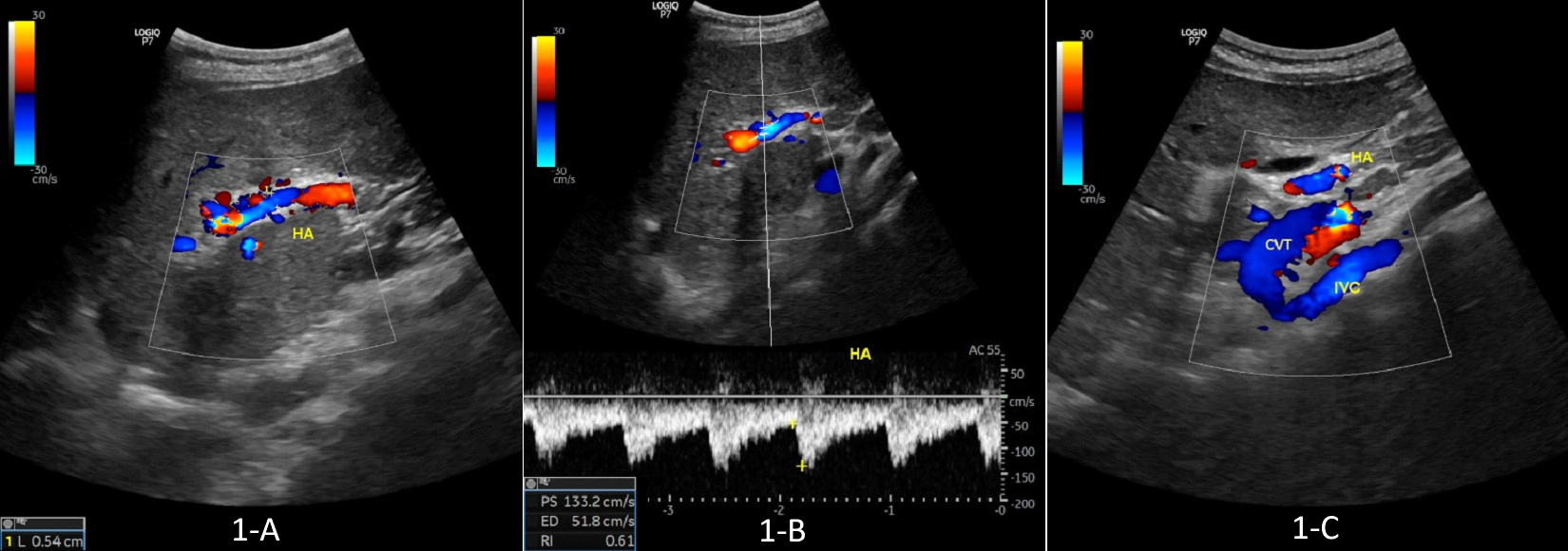
Methods: A 15-year-old female presented with five months of abdominal pain. Her medical history included splenectomy in infancy and repair of an atrial septal defect at 8 months of age. Laboratory evaluation was notable for mildly elevated INR (1.31) and serum ammonia (87 µg/dL), with otherwise normal liver function tests. No esophageal or fundic varices were observed on esophagogastroduodenoscopy (EGD). Abdominal ultrasound demonstrated situs inversus, hepatomegaly, hepatic masses, and an accessory spleen. Doppler ultrasound revealed an absent portal vein, an enlarged hepatic artery, and a venous trunk draining into the IVC, raising concern for an extrahepatic portosystemic shunt. Contrast-enhanced CT confirmed the diagnosis of Abernethy malformation Type Ib, with characteristic hepatic lesions showing arterial enhancement and central scars, consistent with focal nodular hyperplasia. Additional anomalies included IVC duplication with azygos continuation, partial annular pancreas, and hepatic veins draining into the right atrium. The patient remained clinically stable and was scheduled for follow-up. Discussion: Abernethy malformation should be suspected in patients with absent portal vein on Doppler ultrasound, particularly when cavernous transformation is not present. Doppler findings such as a draining venous trunk and prominent hepatic artery may be the first clues to diagnosis. Cross-sectional imaging helps confirm the subtype and identify associated anomalies. Early recognition of abnormal Doppler findings can prompt appropriate cross-sectional imaging and lead to accurate classification of this rare vascular anomaly.
Figure: Figure 1: Abdominal Doppler ultrasound. 1-A: Abdominal Doppler ultrasound of the porta hepatis showing absence of portal vein with multiple tiny venules and dilated hepatic artery (5.4 mm). 1-B: Pulsed Doppler ultrasound showing normal resistive index of the enlarged hepatic artery (0.61). 1-C: Abdominal Doppler ultrasound showing a common venous trunk (CVT) draining into the inferior vena cava (IVC).
Disclosures: Hany Habib indicated no relevant financial relationships. Khaldoun Arabi indicated no relevant financial relationships. Anas Alsolaiman indicated no relevant financial relationships. Samir Haffar indicated no relevant financial relationships.
Hany Habib, MD1, Khaldoun Arabi, MD2, Anas Alsolaiman, MD3, Samir Haffar, MD4. P1800 - Abdominal Pain in a Young Girl With Multiple Congenital Malformations, ACG 2025 Annual Scientific Meeting Abstracts. Phoenix, AZ: American College of Gastroenterology.

.jpg "Hany Habib, MD photo")