Hari Movva, MD1, Shiv R. Patel, MD2, Christo Mathew, MD3, Vikas Burugu, MD1, Jimmy George, MD1, Peeyush Bhargava, 1 1University of Texas Medical Branch, Galveston, TX; 2University of Virginia, Charlottesville, VA; 3University of Texas Medical Branch, Department of Gastroenterology and Hepatology, Galveston, TX Introduction: Hepatic hemangiomas are the most common benign vascular liver lesions and often appear incidentally on imaging, especially MRI. These lesions typically remain asymptomatic and stable in size, though they can enlarge. In patients with cirrhosis, hemangioma behavior may change due to altered hepatic architecture and perfusion. We describe two cases in which hepatic hemangiomas involuted in the setting of cirrhosis.
Case Description/
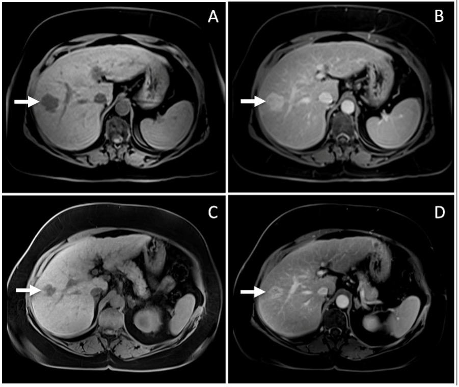
Methods: A 54-year-old woman with untreated hepatitis C–related cirrhosis underwent MRI for hepatocellular carcinoma (HCC) surveillance. Imaging revealed a 3 cm right hepatic lobe lesion consistent with a hemangioma and no suspicious findings. Six months later, follow-up MRI showed the hemangioma had decreased to 2.1 cm. Her MELD-Na score remained stable at 11, and she showed no signs of hepatic decompensation during this interval. Hemangioma size decreased while the liver morphology changed from hepatic steatosis to increased nodularity, caudate hypertrophy, an increased parenchymal heterogeneity with similar overall size.
A 69-year-old man with alcohol-associated cirrhosis initially underwent MRI 10 years ago to evaluate a CT-detected liver lesion. The scan confirmed a 3 cm hemangioma in the right hepatic lobe. Two years later, surveillance MRI revealed the hemangioma had decreased to 2.4 cm. During that time, he developed peripheral edema, and the imaging showed worsening cirrhotic morphology and signs of portal hypertension. Interestingly, his MELD-Na score declined from 12 to 8. Hemangioma size decreased while the liver began with cirrhotic liver morphology with modest nodularity. Interval scan showed increased nodularity and unchanged parenchymal heterogeneity
Discussion: While hepatic hemangiomas are common, few reports describe their behavior in cirrhosis. These cases demonstrate a pattern of hemangioma regression that may reflect progressive hepatic fibrosis and altered vascular flow. Prior studies support this observation: one case series of 21 hemangiomas in 17 cirrhotic patients showed size reduction in one-third of lesions. In cirrhotic livers, distorted architecture may obscure or compress vascular lesions, further complicating radiographic interpretation. MRI remains the preferred modality for accurate monitoring. These cases underscore the importance of considering cirrhotic progression when evaluating changes in hepatic hemangiomas and highlight a need for further investigation into this underrecognized phenomenon.
Figure: Interval decrease in size of hemangioma in patient 1 (A to B), and patient 2 (C to D).
Disclosures: Hari Movva indicated no relevant financial relationships. Shiv Patel indicated no relevant financial relationships. Christo Mathew indicated no relevant financial relationships. Vikas Burugu indicated no relevant financial relationships. Jimmy George indicated no relevant financial relationships. Peeyush Bhargava indicated no relevant financial relationships.
Hari Movva, MD1, Shiv R. Patel, MD2, Christo Mathew, MD3, Vikas Burugu, MD1, Jimmy George, MD1, Peeyush Bhargava, 1. P1732 - Disappearing Acts: Shrinking Hemangiomas in the Setting of Cirrhosis, ACG 2025 Annual Scientific Meeting Abstracts. Phoenix, AZ: American College of Gastroenterology.