Sunday Poster Session
Category: Liver
P1676 - Harnessing Deep Learning for Precision in Liver Tumor Segmentation: A Meta-Analysis of Performance Across Benchmark CT Datasets
Sunday, October 26, 2025
3:30 PM - 7:00 PM PDT
Location: Exhibit Hall

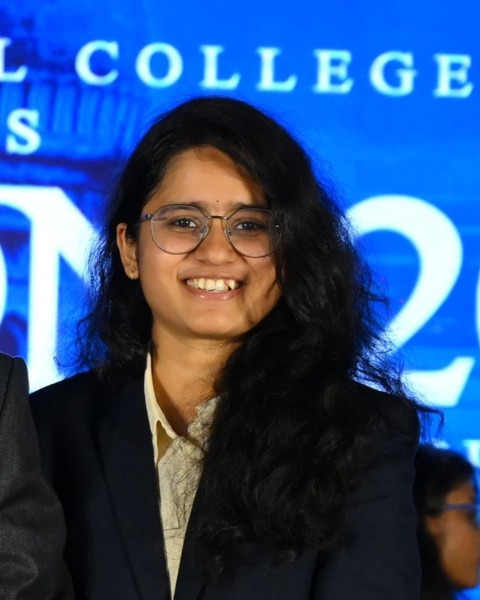
Nishmitha Puchakayala, MBBS (she/her/hers)
Osmania General Hospital and Medical College
Khammam, Telangana, India
Presenting Author(s)
Krishna Teja Vemulaghat, MBBS1, Sahithi Gopu, MBBS2, Bhavana Korlakunta, 3, Nishmitha Puchakayala, MBBS4, Deekshith Ameer Shaik, MBBS5, Balakrishnan Kamaraj, 6
1Osmania General Hospital and Medical College, Siddipet, Telangana, India; 2Osmania General Hospital and Medical College, Godhavarikhani, Telangana, India; 3Osmania General Hospital and Medical College, Aswapuram, Telangana, India; 4Osmania General Hospital and Medical College, Khammam, Telangana, India; 5Osmania General Hospital and Medical College, Hyderabad, Telangana, India; 6Madurai Medical College, Vellore, Tamil Nadu, India
Introduction: Deep learning has emerged as a transformative force in medical imaging. For liver tumors—a significant burden in hepatology—accurate segmentation on CT imaging is critical. Manual segmentation is time-intensive and error-prone; artificial intelligence (AI) offers a promising alternative.
Methods: A comprehensive search of PubMed, Cochrane Library, Embase, and Web of Science was conducted for studies published between May 2017 and April 2024, following the PRISMA guidelines. Quality assessment of these studies was conducted using the CLAIM and QUADAS-2 tools to evaluate applicability and bias. The models in those studies selected for meta-analysis were trained and validated on either the MICCAI LiTS 2017 or 3DIRCADb datasets. These models' reported dice similarity coefficients (DSC) were utilised for meta-analysis. Factors affecting algorithm performance were investigated. Wilcoxon Signed-Rank Test was conducted to compare the distribution among two datasets.
Results: Out of 224 identified studies, 41 were included in the meta-analysis, with 31 models evaluated on the LiTS 2017 dataset and 25 on 3DIRCADb. The top-performing algorithms achieved Dice Similarity Coefficients (DSC) of 0.846 (SD ≈ 0.078, IQR ≈ 0.103) on LiTS 2017 and 0.827 (SD ≈ 0.071, IQR ≈ 0.070) on 3DIRCADb. A Wilcoxon signed-rank test yielded a p-value of 0.064.
Among models tested on both datasets, SADSNet and MAPFUNet performed best. DenseUNet and Spatial UNet outperformed Cascaded UNet and ResUNet, while DCNN and FCN variants surpassed traditional CNNs. Specialized models like CEDRNN and GCN showed strong results but were only evaluated on LiTS 2017.
Discussion: AI-driven segmentation demonstrates high accuracy and reliability, suggesting its readiness for clinical integration in hepatology workflows. With further development and validation, such tools could reduce inter-observer variability, accelerate diagnostic timelines, and support personalized treatment planning in liver cancer management.
Disclosures:
Krishna Teja Vemulaghat indicated no relevant financial relationships.
Sahithi Gopu indicated no relevant financial relationships.
Bhavana Korlakunta indicated no relevant financial relationships.
Nishmitha Puchakayala indicated no relevant financial relationships.
Deekshith Ameer Shaik indicated no relevant financial relationships.
Balakrishnan Kamaraj indicated no relevant financial relationships.
Krishna Teja Vemulaghat, MBBS1, Sahithi Gopu, MBBS2, Bhavana Korlakunta, 3, Nishmitha Puchakayala, MBBS4, Deekshith Ameer Shaik, MBBS5, Balakrishnan Kamaraj, 6. P1676 - Harnessing Deep Learning for Precision in Liver Tumor Segmentation: A Meta-Analysis of Performance Across Benchmark CT Datasets, ACG 2025 Annual Scientific Meeting Abstracts. Phoenix, AZ: American College of Gastroenterology.
