Sari Lada, DO1, Ariel Ahl, MD2, Stella Zaringhalam, MD2, Julie Huynh, MD3, Kumar Desai, MD2 1HCA Healthcare, Woodland Hills, CA; 2HCA Healthcare, Thousand Oaks, CA; 3David Geffen School of Medicine at UCLA, Tarzana, CA Introduction: Extramedullary plasmacytomas (EMPs) are rare plasma cell tumors arising outside bone marrow (~3% of plasma cell disorders). Pancreatic involvement (< 5% of EMPs) mimics pancreatic adenocarcinoma clinically and radiologically. Accurate differentiation is crucial due to significant treatment and prognosis differences. This case highlights pancreatic EMP in a multiple myeloma patient, emphasizing the need for careful evaluation and diagnosis.
Case Description/
Methods: A 77-year-old woman with hypertension, hyperlipidemia, hypothyroidism, anemia, melanoma, and untreated extramedullary plasmacytoma was evaluated for a pancreatic head mass noted on imaging.
Initially presenting with a left breast mass, her exam also revealed pedal edema. Labs showed hemoglobin 11.1 g/dL, hematocrit 33.3%, RBC 3.48 × 10⁶/µL, and elevated MCV (95.7 fL). Mammography identified an oval breast mass, with ultrasound showing suspicious solid lesions. Breast biopsy confirmed a plasma cell neoplasm (CD38/CD138-positive, kappa light chain-restricted).
Further labs revealed an elevated kappa:lambda ratio (4.21) and high kappa free light chains (26.34 mg/L). Bone marrow had 12% plasma cells. PET imaging detected lesions in the forehead, pancreatic head, costovertebral joint, abdominal wall, and diffuse marrow uptake. MRCP characterized an 11 mm hyperenhancing pancreatic head lesion.
Forehead biopsy indicated aggressive kappa-clonal plasma cells (Ki67 >90%). Bone marrow biopsy showed 25% kappa-restricted plasma cells, confirming multiple myeloma. FISH revealed high-risk cytogenetics (gain 1q, loss 1p, monosomy 13, partial 5’IGH deletion, loss 16q).
EUS showed a 12 × 15 mm hypoechoic pancreatic mass. Fine-needle biopsy confirmed plasmacytoma (50% cellularity, CD138-positive, IgG-positive, Ki67 proliferation 50%). The patient began systemic treatment with daratumumab, lenalidomide, aspirin, acyclovir, and dexamethasone. Discussion: Extramedullary plasmacytoma (EMP) of the pancreas is rare and often appears similar to pancreatic adenocarcinoma, typically causing jaundice and abdominal pain. Most commonly found in the head of the pancreas, EMPs occur more frequently in patients with multiple myeloma and usually indicate aggressive underlying disease. Fortunately, EMPs tend to have a better prognosis than pancreatic adenocarcinoma, highlighting the importance of timely biopsy and immunohistochemistry. Early and accurate diagnosis can lead to more effective treatment, reducing unnecessary surgeries and significantly improving outcomes.
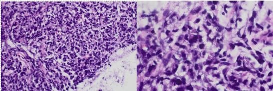
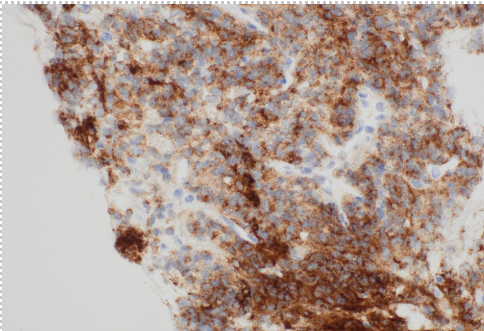
Figure: Pancreatic biopsy shows predominant population of atypical plasma cells that are CD138 positive: Hematoxylin and Eosin staining, magnification x 40 and x100.
Disclosures: Sari Lada indicated no relevant financial relationships. Ariel Ahl indicated no relevant financial relationships. Stella Zaringhalam indicated no relevant financial relationships. Julie Huynh indicated no relevant financial relationships. Kumar Desai indicated no relevant financial relationships.
Sari Lada, DO1, Ariel Ahl, MD2, Stella Zaringhalam, MD2, Julie Huynh, MD3, Kumar Desai, MD2. P1429 - A Case of Extramedullary Plasmacytoma Involving the Pancreas, ACG 2025 Annual Scientific Meeting Abstracts. Phoenix, AZ: American College of Gastroenterology.

 photo")