Louisiana State University Health Sciences Center New Orleans New Orleans, LA
Nikki Arceneaux, MD1, Blake Savoie, MD1, Stephen Landreneau, MD, FACG2 1Louisiana State University Health Sciences Center New Orleans, New Orleans, LA; 2LSU Health Sciences Center New Orleans, New Orleans, LA Introduction: Liposarcoma of the abdomen is a rare malignant tumor of adipocytes. While most commonly arising in the deep soft tissues of the extremities, cases have been reported in the stomach and retroperitoneum. Symptoms are often nonspecific and typically do not present until the tumor reaches a large size. The etiology remains unclear, but potential risk factors include prior radiation exposure, lymphatic system trauma, and toxic chemical exposure. Diagnosis is confirmed by biopsy with fluorescence in situ hybridization (FISH) testing for amplification of the mouse double minute 2 (MDM2). We present a case of abdominal liposarcoma diagnosed by endoscopic ultrasound-guided fine needle biopsy (EUS-FNB).
Case Description/
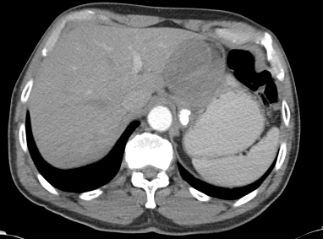
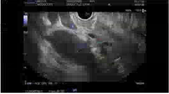
Methods: A 64-year-old male with a past medical history of sarcoidosis with cutaneous, cardiac, and pulmonary involvement presented to GI clinic for evaluation of a perigastric/gastric mass discovered incidentally on computed tomography (CT). Imaging showed a heterogeneous mass adjacent to the stomach and left hepatic lobe measuring 6.1 x 6.5 cm. He denied abdominal symptoms such as pain or nausea. EUS showed two masses in the gastrohepatic space measuring 68 mm x 82 mm and 50 mm x 48 mm. It was unclear if these masses were contiguous. Fine needle biopsies and histologic sections demonstrated a diffusely sclerotic and hyalinized mass with scattered adipocytic tissue, mixed inflammation, and rare hyperchromatic atypical cells. No clear mitotic activity was present. MDM2 FISH testing was positive for amplification of the MDM2 gene. These findings support a diagnosis of well-differentiated liposarcoma (WDLS), sclerosing type. He was referred to medical and surgical oncology for treatment planning. Discussion: Liposarcomas are typically classified into four subtypes based on their histological and molecular characteristics: well-differentiated liposarcoma (WDLS), dedifferentiated liposarcoma, myxoid and/or round cell, and pleomorphic. Mature adipocytes and fibrous stroma with atypical nuclei histologically characterize WDLS. It is classified into three subtypes: lipoma-like, sclerosing, and inflammatory. The sclerosing subtype, as seen in this patient, features dense fibrous stroma with minimal lipogenic tissue. MDM2 amplification can help differentiate WDLS from benign lipomas. Surgical resection with negative margins remains the cornerstone of treatment. Although WDLS has a favorable prognosis, it carries a high risk of local recurrence, necessitating regular surveillance imaging.
Figure: Axial contrast-enhanced abdominal CT image showing a heterogeneous mass adjacent to the stomach and left hepatic lobe measuring approximately 6.1 x 6.5 cm.
Figure: Endoscopic ultrasound of a mass within the gastrohepatic space measuring approximately 68 mm by 82 mm.
Disclosures: Nikki Arceneaux indicated no relevant financial relationships. Blake Savoie indicated no relevant financial relationships. Stephen Landreneau indicated no relevant financial relationships.
Nikki Arceneaux, MD1, Blake Savoie, MD1, Stephen Landreneau, MD, FACG2. P1427 - Abdominal Liposarcoma Diagnosed by Endoscopic Ultrasound-Guided Fine Needle Biopsy, ACG 2025 Annual Scientific Meeting Abstracts. Phoenix, AZ: American College of Gastroenterology.

 photo")