MedStar Georgetown/Washington Hospital Center Washington, DC
Bara Abujaber, MBBS1, Nour Abu Irhayem, MBBS2, Kenan Alrejjal, MBBS1, Alireza Meighani, MD3 1MedStar Georgetown/Washington Hospital Center, Washington, DC; 2MedStar Georgetown/ Washington Hospital Center, Washington, DC; 3MedStar Georgetown University Hospital, Washington, DC Introduction: Clostridioides difficile infection (CDI) is a leading cause of healthcare-associated diarrhea. Its typical endoscopic findings include erythema, edema, and pseudomembranes. Deep colonic ulcerations are exceedingly rare and may mimic ischemic colitis or inflammatory bowel disease (IBD), often resulting in misdiagnosis or delayed treatment. We present a case of CDI with extensive colonic ulceration and an initially negative stool test, underscoring the diagnostic value of colonoscopy in atypical presentations.
Case Description/
Methods: A 70-year-old woman with Hodgkin lymphoma, supraventricular tachycardia, and hypothyroidism was hospitalized for type 2 atrioventricular block. During admission, she developed hospital-acquired pneumonia treated with meropenem, followed by profuse diarrhea. CDI was confirmed and resolved after a 10-day course of oral vancomycin. Eight days post-treatment, she received ceftriaxone for a urinary tract infection and developed recurrent diarrhea. Initial CDI stool testing was negative. She had no prior GI complaints or IBD history. Due to persistent symptoms, CT angiography was performed, showing pancolitis concerning for ischemic or inflammatory colitis. Colonoscopy revealed moderate-to-severe colitis with diffuse shallow and deep ulcerations, an unusual CDI finding. Viral testing, including CMV and HSV, was negative. Biopsies demonstrated acute inflammation without chronic changes or ischemic features. Given her recent CDI and antibiotic exposure, repeat stool testing confirmed recurrent CDI. She was treated with pulsed oral vancomycin leading to complete resolution within three days. Discussion: This case challenges the conventional presentation of CDI, demonstrating that deep colonic ulcerations can be a primary CDI manifestation. Colonoscopy was key in clarifying the diagnosis when imaging was inconclusive. While CDI and ischemic colitis share features, the absence of watershed distribution grossly and ischemic changes on biopsy both strongly favor CDI. IBD was unlikely given the lack of prior GI symptoms, chronic inflammation, or granulomas, with a clinical course tied to recent antibiotics and CDI recurrence. Ultimately, the strongest evidence for CDI was the rapid symptom resolution with targeted treatment. Recognizing deep ulcers as a CDI feature is crucial to prevent misdiagnosis. This case features the limitations of initial single negative CDI tests, the need for repeat testing in high-risk patients, and the importance of early colonoscopy in atypical cases.
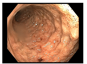
Figure: Figure 1. Colonoscopy demonstrating Ulcers at Sigmoid colon.
Figure: Figure 2. Ulcers associated with acute inflammation with cryptitis, crypt abscesses and mucin with neutrophilic exudate.
Disclosures: Bara Abujaber indicated no relevant financial relationships. Nour Abu Irhayem indicated no relevant financial relationships. Kenan Alrejjal indicated no relevant financial relationships. Alireza Meighani indicated no relevant financial relationships.
Bara Abujaber, MBBS1, Nour Abu Irhayem, MBBS2, Kenan Alrejjal, MBBS1, Alireza Meighani, MD3. P1360 - Beyond Pseudomembranes: Deep Colonic Ulcers in Clostridioides difficile Infection, ACG 2025 Annual Scientific Meeting Abstracts. Phoenix, AZ: American College of Gastroenterology.

 photo")