Reema Wagh, BS, David Karow, MD, Jana G. Hashash, MD, MSc, FACG Mayo Clinic, Jacksonville, FL Introduction: The spleen slightly atrophies naturally as people age, but this shrinkage can be caused by many other factors including infections such as malaria and HIV, autoimmune diseases such as systemic lupus erythematosus, malnutrition, and other diseases such as celiac disease, Whipple’s disease and inflammatory bowel diseases (IBD). Splenic atrophy may lead to increase risk of infection and impairment in defense against infections, as well as abnormalities in blood counts (thrombocytosis and leukocytosis).
Case Description/
Methods: A 29-year-old female with ileocolonic Crohn’s disease of 7 year duration, who is currently maintained on upadacitinib 30 mg daily, presents to establish care in our clinic. She has been having symptoms of active disease characterized by non-bloody loose stools occurring 3 times per day and diffuse abdominal pain. Prior medical therapy includes mesalamine, ozanimod, and vedolizumab.
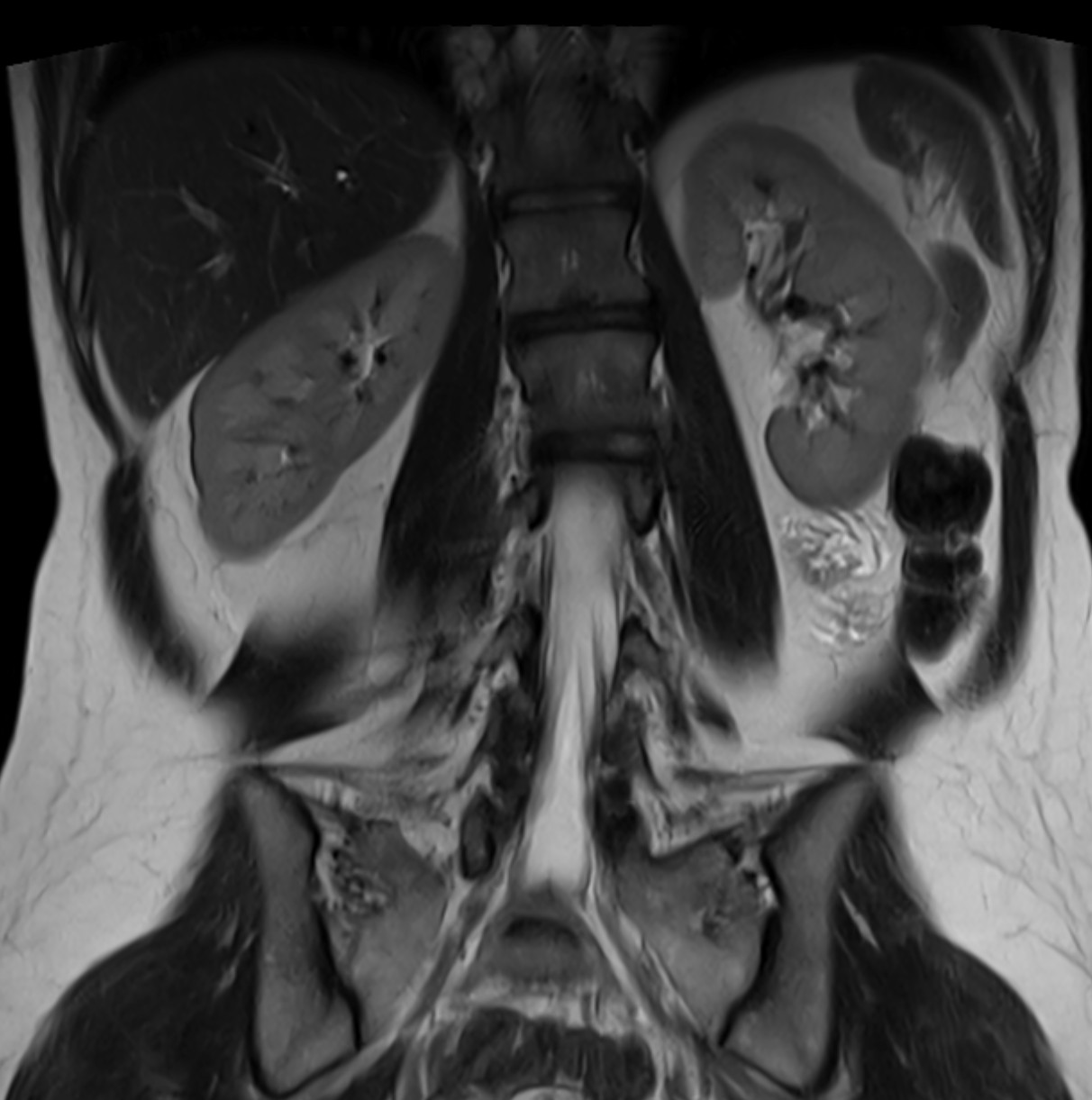
To further evaluate her abdominal pain and to assess for disease extemt, cross sectional imaging in the form of a magnetic resonance enterography (MRE) was performed and showed mild to moderate wall thickening and enhancement in the right and transverse colon; and to a lesser degree in the left colon (descending colon to rectum). The small bowel appeared normal. Interestingly, the spleen appeared atrophic and measured 51 cm3 in size (Figure 1). Normal splenic volume typically ranges from 107.2 to 314.5 cm³, with a mean of 214.6 cm³ in healthy individuals. Discussion: The long standing inflammation in patients with chronic IBD, especially patients with ulcerative colitis, can cause splenic atrophy and hyposplenism. There are varying statistics reported regarding patients who have IBD and atrophic spleen, but this is not uncommon, occurring in 30-37% of patients with UC and to a lesser degree, 5% of patients with Crohn’s disease. Hyposplenism can cause a greater susceptibility to infections such as Streptococcus pneumoniae and Haemophilus influenzae. Vaccinations and at times antibiotic prophylaxis are recommended to reduce the risk of infection.
The reasons behind an atrophic spleen in patients with IBD is multifactorial and likely related to inflammatory burden, cytokine effects, and possible intravascular thrombosis, as well as malabsorption and nutrient deficiencies.
Attention to spleen size on imaging studies of patients with IBD is crucial as spleen size is associated with increased infection risk and disease complication.
Figure: Image taken from the Coronal T2 weighted HASTE images performed on a 3 Tesla Siemens Vida scanner. Spleen dimensions: 6.4 cm cranial caudal x 3.8 cm transverse x 2.1 cm anterior posterior.
Disclosures: Reema Wagh indicated no relevant financial relationships. David Karow indicated no relevant financial relationships. Jana Hashash: BMS – Ad Board.
Reema Wagh, BS, David Karow, MD, Jana G. Hashash, MD, MSc, FACG. P1272 - Atrophic Spleen: What Could It Be?, ACG 2025 Annual Scientific Meeting Abstracts. Phoenix, AZ: American College of Gastroenterology.