University of Texas at Austin Dell Medical School Austin, TX
Award: ACG Presidential Poster Award
Jonathan Pham, MD, Linda A. Feagins, MD University of Texas at Austin Dell Medical School, Austin, TX Introduction: Objective assessment of disease activity is a critical component of the treat-to-target paradigm in Crohn’s disease (CD) management. Colonoscopy remains the gold standard; however, when endoscopic small bowel evaluation is limited due to strictures, we have historically turned to CT/MR enterography, which has its own limitations with cost, availability, and need for oral contrast preparation. Intestinal ultrasound (IUS) offers a non-invasive, cost-effective technique for point-of-care assessment of disease activity proximal to strictures and may aid in therapeutic decision-making. We present four cases of patients with CD who underwent outpatient colonoscopy with suspected or new strictures preventing proximal evaluation, and IUS was used same-day to evaluate ileal disease activity proximal to the stricture.
Case Description/
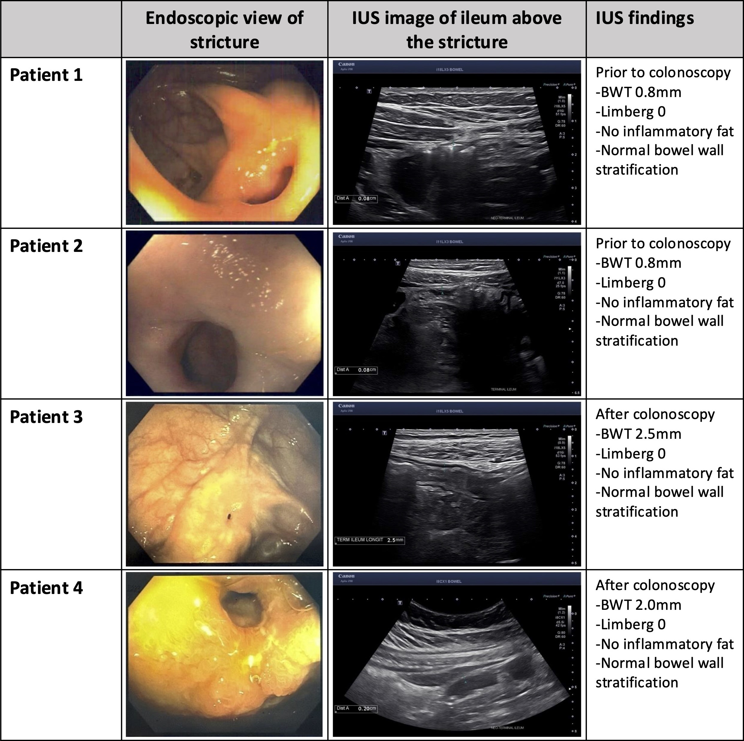
Methods: Four patients with ileocolonic CD underwent outpatient colonoscopy (Table 1). Three patients had prior ileocolonic resection with ileocolonic anastomosis (ICA) and one had a native ileocecal valve (ICV). Two patients had strictures at the ICA, one proximal to the ICA, and one at the ICV, all preventing endoscopic evaluation proximal to the stricture. IUS was performed before colonoscopy in two patients and after colonoscopy in two patients, which showed no evidence of active disease in all 4 cases (Figure 1). Bowel preparation did not prevent visualization of the ileum. In three patients, the mucosa proximal to the stricture was blindly biopsied. Pathology showed normal small bowel mucosa in two patients and minimally active ileitis in one patient. In the fourth patient who had a stricture proximal to the ICA, biopsies were unable to be obtained across the stricture. One patient had an MR enterography that was performed one-month post-procedure, which also confirmed no active ileal disease proximal to the stricture. Discussion: In this case series, four patients with CD who had ICV or ICA strictures underwent colonoscopy and same-day IUS. Targeted ultrasound proximal to the stricture showed inactive disease in each case, consistent with biopsies taken through the stricture and/or with MR enterography performed post-procedure. This highlights the potential of IUS as an adjunctive tool to guide real-time therapeutic decision-making by assessing disease activity proximal to strictures, especially when encountered unexpectedly during colonoscopy.
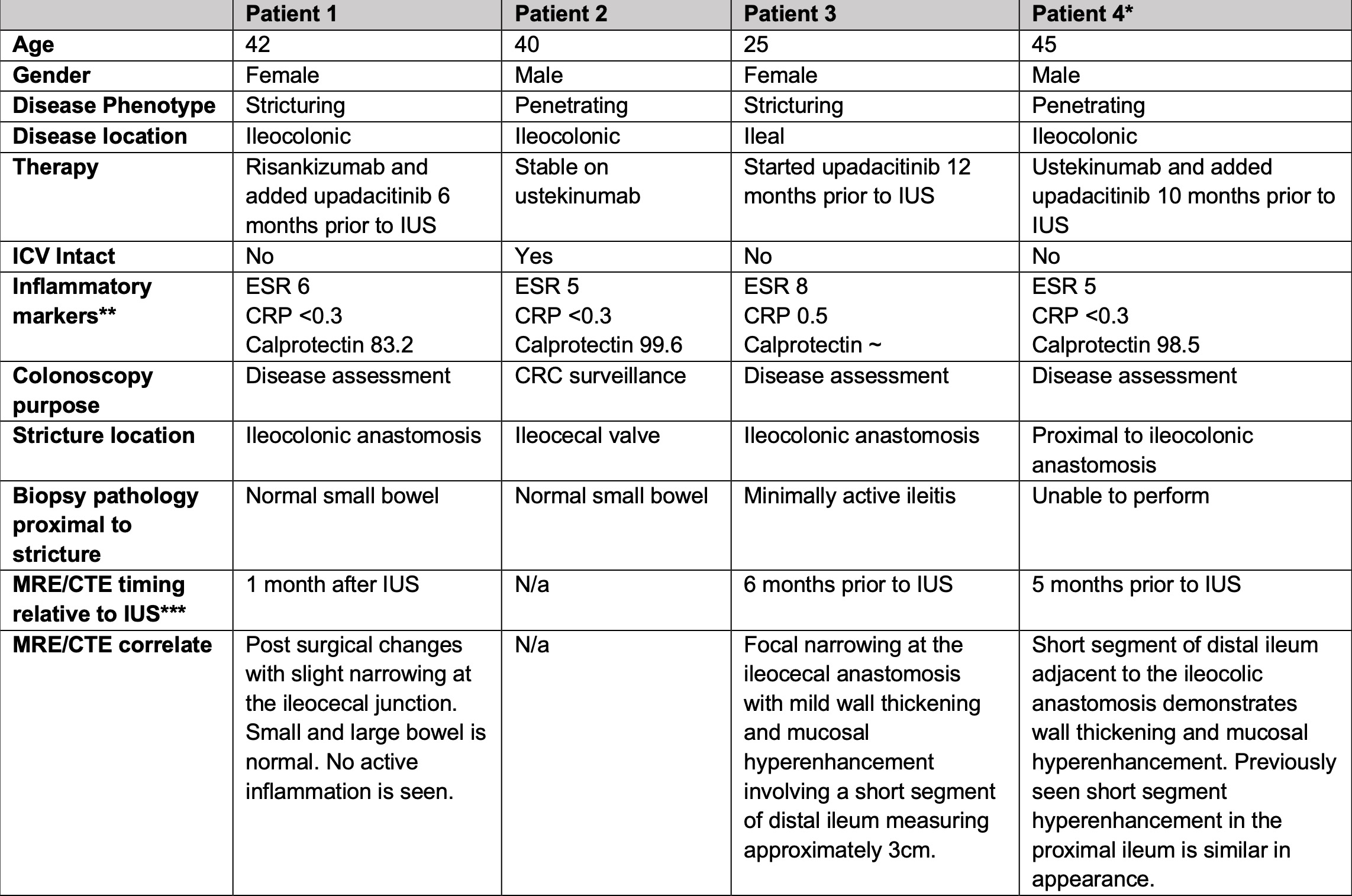
Figure: Table 1: Details of 4 cases of patients undergoing colonoscopy with same-day IUS *Patient had neo-terminal ileum edema and mild ulceration distal to the ileocolonic anastomosis **Inflammatory markers are presented +/- 3 months from colonoscopy. Erythrocyte sedimentation rate (ESR): normal 0-20mm/hr, C-reactive protein (CRP): normal <0.5mg/dL *** MRE/CTE results +/- 6 months from colonoscopy/IUS are presented
Figure: Figure 1: Endoscopic evaluation of intestinal strictures and intestinal ultrasound evaluation of ileal disease activity proximal to the stricture in four patients
Disclosures: Jonathan Pham indicated no relevant financial relationships. Linda Feagins: Corevitas – Clinical trial participation. Takeda – Clinical trial participation.
Jonathan Pham, MD, Linda A. Feagins, MD. P1227 - Intestinal Ultrasound for Ileal Disease Activity Assessment in Crohn’s Patients With Strictured Ileocecal Valves or Ileocolonic Anastomoses, ACG 2025 Annual Scientific Meeting Abstracts. Phoenix, AZ: American College of Gastroenterology.