Memorial Health University Medical Center Savannah, GA
Helen Paglia, DO1, Marco Noriega, MD, MPH2, Kinnari Kher, MD2 1Memorial Health University Medical Center, Savannah, GA; 2Mount Auburn Hospital, Harvard Medical School, Cambridge, MA Introduction: Upper gastrointestinal bleeding (UGIB) is a common and potentially life-threatening condition, with peptic ulcer disease being the most frequent cause. Malignancy accounts for a small proportion of UGIB cases, and extranodal involvement by non-Hodgkin’s lymphoma is particularly rare. Diffuse large B-cell lymphoma (DLBCL), the most common subtype of non-Hodgkin’s lymphoma, can involve extranodal sites including the gastrointestinal tract. We present a rare case of UGIB due to DLBCL involving the stomach, liver, gallbladder, and inferior vena cava (IVC).
Case Description/
Methods: An 80-year-old man with no prior gastrointestinal history presented with two days of coffee-ground emesis, fatigue, melena, and lower extremity edema. He was hemodynamically stable but profoundly anemic with a hemoglobin of 4.9 g/dL. Initial concern for alcohol-related UGIB prompted abdominal imaging, which revealed a large heterogenous mass in the right hepatic lobe, abutting the gallbladder, and a 5.4 x 4.7 cm tumor thrombus in the IVC. Upper endoscopy revealed a large, clean-based 3 cm gastric ulcer with rolled margins and coffee-ground material in the base. An endoscopic clip was attempted but could not be deployed due to mucosal tethering. Biopsy of the liver mass confirmed DLBCL evolving from follicular lymphoma. Recurrent bleeding necessitated repeat endoscopy, during which a visible vessel and adherent clot were treated with bipolar cautery, epinephrine injection, and hemostatic spray. Discussion: Although peptic ulcer disease remains the most common cause of UGIB, malignancy must be considered, especially in patients with constitutional symptoms or unusually large gastric ulcers. Ulcers 3 cm or larger should prompt cross-sectional imaging to evaluate for lymphoma as a potential underlying cause. Endoscopic hemostasis in malignant ulcers poses technical challenges due to tumor neovascularization and mucosal tethering, often preventing adequate clip deployment. In such cases, adjunct therapies like hemostatic spray are valuable. This case highlights the importance of considering lymphoma in the differential diagnosis of UGIB and adapting endoscopic strategy to the unique challenges posed by malignancy-related bleeding.
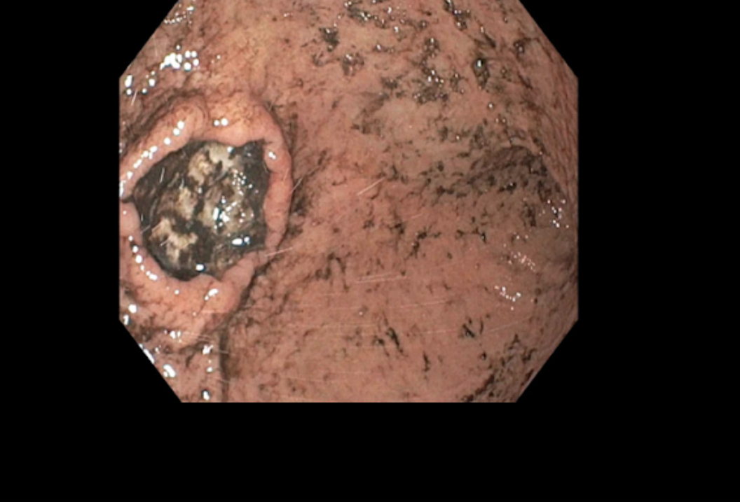
Figure: Image III: Ulcer on greater curvature with coffee ground material in the ulcer base.
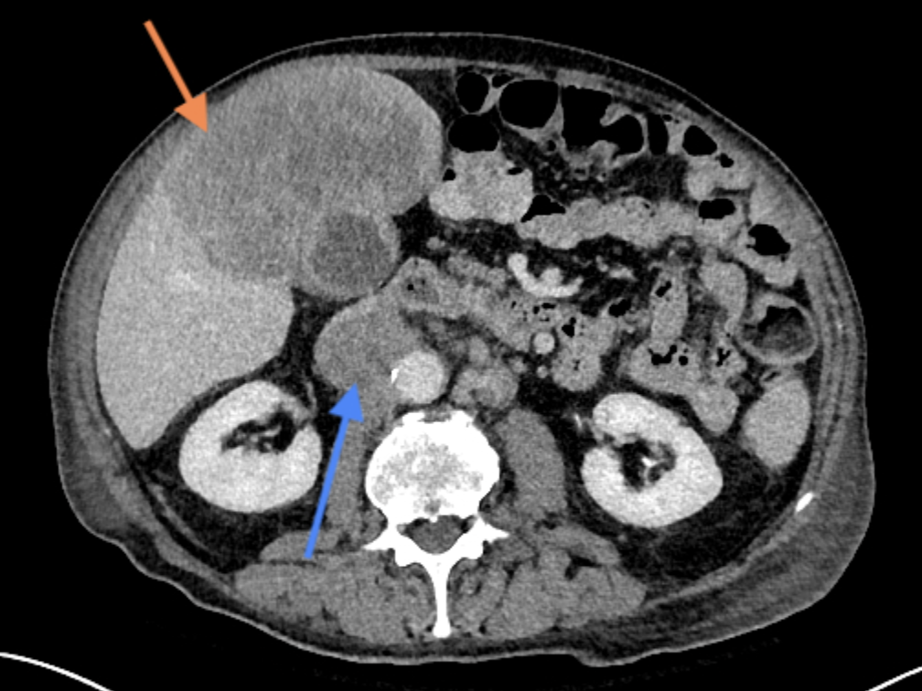
Figure: Image I: CT abdomen transverse section. Orange arrow: mass surrounding the gallbladder. Blue arrow: IVC tumor thrombus.
Disclosures: Helen Paglia indicated no relevant financial relationships. Marco Noriega indicated no relevant financial relationships. Kinnari Kher indicated no relevant financial relationships.
Helen Paglia, DO1, Marco Noriega, MD, MPH2, Kinnari Kher, MD2. P1005 - Diffuse Large B-Cell Lymphoma Presenting as Upper Gastrointestinal Bleeding: A Case Report, ACG 2025 Annual Scientific Meeting Abstracts. Phoenix, AZ: American College of Gastroenterology.


 photo")