University of Texas Medical Branch League City, TX
Christo Mathew, MD1, George Wahba, MD2, Jeffrey H.. Lee, MD, MPH3 1University of Texas Medical Branch, League City, TX; 2MD Anderson Cancer Center, Houston, TX; 3The University of Texas MD Anderson Cancer Center, Houston, TX Introduction: Duodenocaval fistulas are an exceptionally rare complication, often associated with malignancy, trauma, or iatrogenic injury. Timely recognition is critical given the risk of catastrophic hemorrhage.
Case Description/
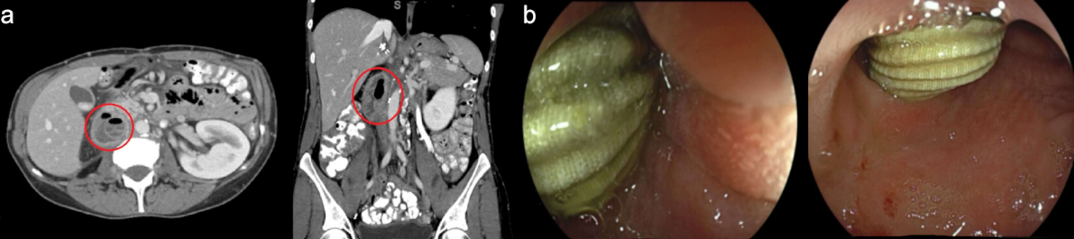
Methods: A 44-year-old woman with recurrent leiomyosarcoma of the inferior vena cava (IVC) had previously undergone surgical resection and IVC graft placement. Her postoperative course was complicated by retroperitoneal fluid collections requiring multiple rounds of percutaneous drainage. She later developed a new, complex, gas-containing collection projecting into the pancreaticoduodenal groove, which was concerning for an abscess. Percutaneous drainage was deemed high risk given the location, and the patient was referred for endoscopic ultrasound (EUS)-guided drainage. During initial endoscopic evaluation, a gastroscope was advanced into the second portion of the duodenum, where a synthetic graft was unexpectedly visualized protruding into the duodenal lumen. This was identified as a Dacron IVC graft. The finding was consistent with a duodenocaval fistula resulting from graft erosion into the duodenum. The endoscopic procedure was terminated, and surgical consultation was obtained. The patient subsequently underwent exploratory laparotomy with graft explantation and duodenal repair. She was discharged in stable condition without complications. Discussion: This case highlights a rare but serious complication of vascular graft placement. Duodenocaval fistulas may present subtly and may be diagnosed incidentally. Endoscopic evaluation, even when performed for other indications, may play a crucial role in early detection. Prompt multidisciplinary coordination is essential to reduce morbidity.
Figure: Figure 1: Computed tomography abdomen of a large complex, gas-containing fluid collection projecting into the pancreaticoduodenal groove, suspicious for abscess (a). Endoscopic view of the synthetic graft was seen emanating into the lumen of the second portion of the proximal duodenum (b).
Disclosures: Christo Mathew indicated no relevant financial relationships. George Wahba indicated no relevant financial relationships. Jeffrey Lee indicated no relevant financial relationships.
Christo Mathew, MD1, George Wahba, MD2, Jeffrey H.. Lee, MD, MPH3. P0881 - Duodenocaval Fistula: Recognizing Unexpected Etiologies with Endoscopy, ACG 2025 Annual Scientific Meeting Abstracts. Phoenix, AZ: American College of Gastroenterology.