Alexander F. Siegel, MD1, Jennifer Gamache, MD2, Aun R. Shah, MBBS, MRCP3, Kyaw Min Tun, DO1 1Creighton University Medical Center, Omaha, NE; 2Creighton University, Omaha, NE; 3CHI Health Creighton University Medical Center, Omaha, NE Introduction: Gastric melanomas are considered uncommon with only a handful of published cases in the literature. We present a case of biopsy-proven cutaneous melanoma that had metastasized to the gastric body in a patient presenting with non-specific gastrointestinal (GI) symptoms and iron deficiency anemia (IDA).
Case Description/
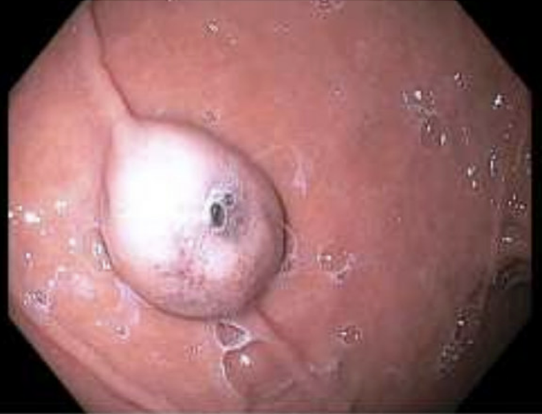
Methods: A 72-year-old female with a history of IDA presented with one week history of left lower quadrant abdominal pain. CT imaging of the abdomen showed nodules in the pelvic mesentery, omentum, gallbladder, retroperitoneal soft tissues and left renal vein, raising concern for metastatic disease of unknown origin. Gastroenterology was initially consulted to help work up the etiology of IDA in this patient who had never undergone upper or lower endoscopy. Colonoscopy demonstrated non-bleeding external hemorrhoids and sigmoid diverticulosis. Upper endoscopy revealed a large polypoid mass on the anterior stomach wall and a non-bleeding gastric ulcer with an adjacent submucosal nodule located in the fundus. Biopsy of the ulcer confirmed malignant melanoma with diagnosis supported by immunohistochemical stains for SOX10 and Melan-A. Three months after discharge the patient’s hemoglobin had increased from 7.2 g/dL to 10.4 g/dL and serum iron increased from 11ug/dL to 22 ug/dL. Without any further hospital admissions for GI issues, it was concluded that patient’s IDA was due to the gastric ulcer and associated submucosal nodule. The patient is currently receiving immunotherapy with Nivolumab/Ipilimumab for melanoma. Discussion: Gastric metastatic disease is relatively uncommon compared to other locations within the GI tract. However, a retrospective observational study analyzing over 400 cases of malignant gastric tumors found melanoma to be the most frequent primary cancer to metastasize to the stomach accounting for 29.6% of cases. Despite this, the majority of malignant melanomas discovered within the GI tract are found within the hepatobiliary system, peritoneum, small bowel and colon. Endoscopic evaluation of gastric malignant melanomas often reveals ulcerated, polypoid, or volcanoid lesions, similar to other metastatic gastric lesions. Thus, histopathological confirmation is essential for accurate diagnosis and is achieved by staining for markers such as SOX10, Melan-A, and HMB-45. This case highlights the importance of considering gastric involvement in patients with a history of melanoma presenting with nonspecific GI symptoms.
Figure: Endoscopic image of gastric melanoma found on the anterior wall of the stomach
Figure: H&E demonstrating evidence of chronic gastritis, focal ulceration and proliferation of neoplastic cells (left). Positive SOX-10 immunohistochemical staining of gastric biopsy consistent with melanoma diagnosis (right).
Disclosures: Alexander Siegel indicated no relevant financial relationships. Jennifer Gamache indicated no relevant financial relationships. Aun Shah indicated no relevant financial relationships. Kyaw Min Tun indicated no relevant financial relationships.
Alexander F. Siegel, MD1, Jennifer Gamache, MD2, Aun R. Shah, MBBS, MRCP3, Kyaw Min Tun, DO1. P0870 - Skin to Stomach: An Uncommon Manifestation of Malignant Melanoma, ACG 2025 Annual Scientific Meeting Abstracts. Phoenix, AZ: American College of Gastroenterology.