Anudeep Jala, DO1, Mansi Sheth, DO2, Daniel Moodey, DO3, Sachin Prasad, DO4, Jason John, DO5, Seth Lipshutz, DO6, Wei Jiang, MD7, Richard Walters, DO8, Drew Chiesa, DO8 1Jefferson Health, Voorhees, NJ; 2Jefferson Torresdale Hospital, Bridgewater, NJ; 3Jefferson Health, Somerdale, NJ; 4Jefferson Health, Blackwood, NJ; 5Jefferson Health, Stratford, NJ; 6Jefferson Health, Cherry Hill, NJ; 7Thomas Jefferson University Hospital, Philadelphia, PA; 8Jefferson Health, Sewell, NJ Introduction: Esophageal cancer comprises several subtypes, the most common being squamous cell carcinoma and adenocarcinoma. Esophageal sarcoma, however, is a rare and aggressive malignancy, accounting for less than 1% of all sarcomas. Due to its rarity, the diagnosis and management of esophageal sarcoma remain challenging and often require a multidisciplinary approach. We present a unique case of esophageal sarcoma presenting with progressive dysphagia.
Case Description/
Methods: A 68-year-old male with a medical history of alcohol use disorder, gastroesophageal reflux disease (GERD), and hypertension presented with a three-week history of nausea, vomiting, hematemesis, and progressive dysphagia with regurgitation of undigested food. In that time, he did report difficulty with tolerating oral intake. Of note, he had never undergone prior esophagogastroduodenoscopy (EGD). Computed tomography (CT) imaging of the chest, abdomen, pelvis with contrast revealed esophageal wall thickening in the mid-to-distal esophagus.
Subsequent EGD revealed an ulcerated, fungating mass involving the middle third of the esophagus and extending into the lower third, causing partial luminal obstruction. A fully covered self-expanding metal stent (FCSEMS) was placed to relieve the obstruction. Immunohistochemical staining of the mass demonstrated tumor cells positive for AE1/AE3 and Cam5.2. Stains were negative for CK7, CK20, CK5/6, p40, SMA, desmin, myogenin, CD117, DOG1, CD34, SOX10, CD45, CD3, CD20, PAX5, CD30, and CD68. The Ki-67 proliferative index was elevated at 70-80%, indicating high-grade malignancy. These findings were consistent with poorly differentiated sarcoma, favoring primary esophageal sarcoma. Discussion: Management of esophageal sarcoma initially involved surgical resection, with esophagectomy for localized esophageal sarcomas. Due to its aggressive nature and high risk of metastasis, a multidisciplinary approach is often required with a combination of systemic chemotherapy and radiation therapy. This case emphasized the importance of maintaining a high index of suspicion for esophageal sarcoma in patients with progressive dysphagia, regurgitation, or signs of upper gastrointestinal obstruction. Prompt diagnosis and intervention are necessary for improving outcomes in this rare malignancy.
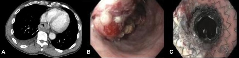
Figure: Figure 1: (A) CT chest, abdomen, and pelvis with contrast demonstrating esophageal wall thickening, (B-C) Ulcerating mass of esophagus with placement of FCSEMS stent.
Disclosures: Anudeep Jala indicated no relevant financial relationships. Mansi Sheth indicated no relevant financial relationships. Daniel Moodey indicated no relevant financial relationships. Sachin Prasad indicated no relevant financial relationships. Jason John indicated no relevant financial relationships. Seth Lipshutz indicated no relevant financial relationships. Wei Jiang indicated no relevant financial relationships. Richard Walters indicated no relevant financial relationships. Drew Chiesa indicated no relevant financial relationships.
Anudeep Jala, DO1, Mansi Sheth, DO2, Daniel Moodey, DO3, Sachin Prasad, DO4, Jason John, DO5, Seth Lipshutz, DO6, Wei Jiang, MD7, Richard Walters, DO8, Drew Chiesa, DO8. P0764 - Esophageal Sarcoma: A Rare Entity Presenting as Progressive Dysphagia, ACG 2025 Annual Scientific Meeting Abstracts. Phoenix, AZ: American College of Gastroenterology.

.jpeg.jpg "Anudeep Jala, DO (he/him/his) photo")