Texas Tech University Health Sciences Center Lubbock, TX
Zeyad Elharabi, MBBS, MS, Romelia A. Barba Bernal, MD, Jowana Saba, MD, Hakan Akin, MD Texas Tech University Health Sciences Center, Lubbock, TX Introduction: Bullous pemphigoid (BP) is an autoimmune subepidermal blistering disorder, primarily affecting the elderly. It is caused by autoantibodies against hemidesmosomal proteins (BP180 and BP230), leading to tense bullae formation, usually on the skin of the trunk and extremities. The involvement of mucous membranes is atypical, but when present, it is generally limited to the oral mucosa. It rarely affects other mucosal surfaces. We present a case of a 77-year-old man with bullous pemphigoid associated with pharyngeal and upper esophageal involvement but no oral mucosal lesions. He was also found to have sigmoid colon adenocarcinoma, raising suspicion for paraneoplastic bullous pemphigoid.
Case Description/
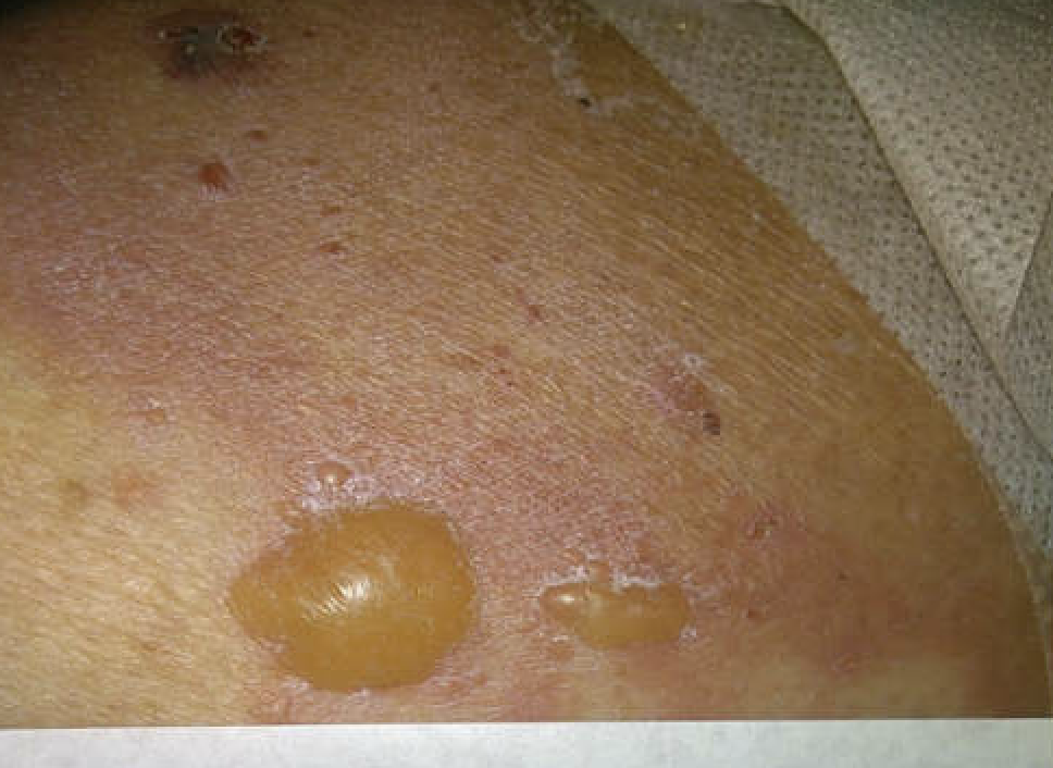
Methods: A 77-year-old man with a past medical history of type 2 diabetes mellitus, hypertension, and cerebrovascular accident presented with a four-weeks history of diarrhea and generalized blisters. He described the blisters as itchy, and he reported that they started on the skin of the abdomen and later spread all over his body. On examination, tense bullae and superficial erosions were noted on his trunk, bilateral upper extremities, and bilateral lower extremities. There were no lesions on the oral mucosa and genitalia. Dermatology was consulted, and punch biopsies were obtained. Histopathology showed changes consistent with bullous pemphigoid. Direct immunofluorescence demonstrated linear IgG and C3 deposition along the basement membrane.
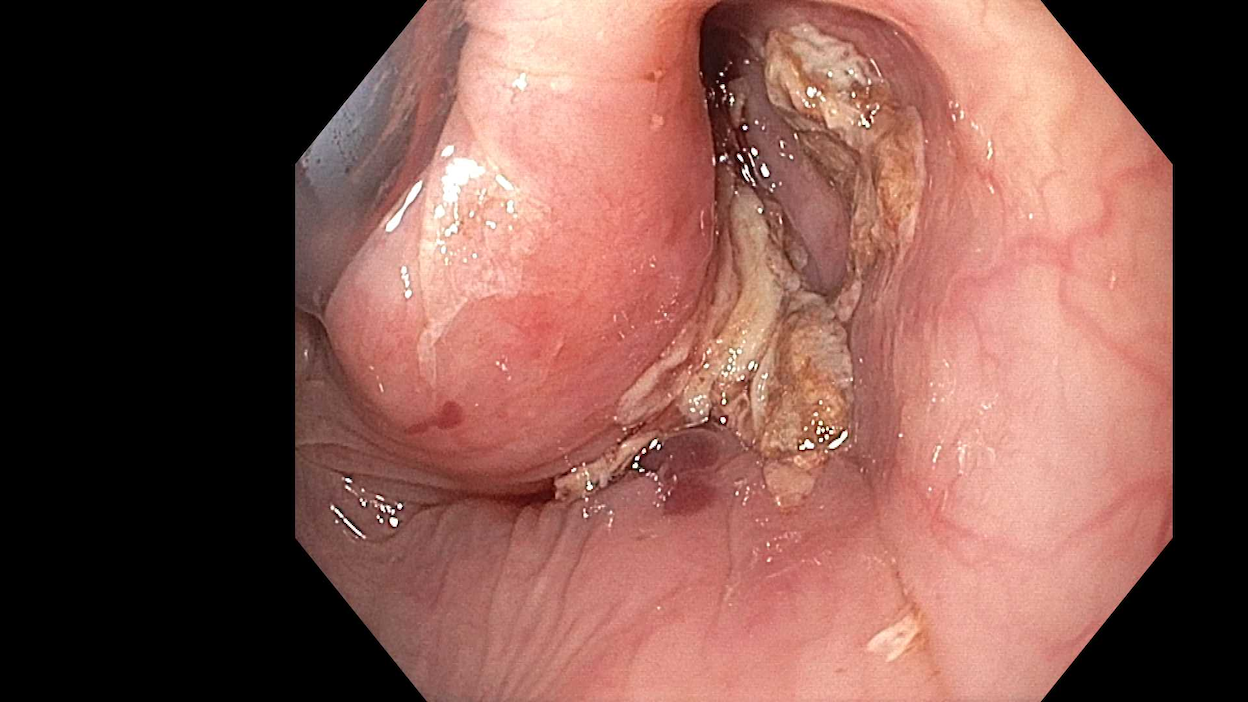
During hospitalization, the patient developed dysphagia. Esophagogastroduodenoscopy revealed ulceration in the hypopharynx and upper esophagus and hemorrhagic bullae in the hypopharynx. Biopsies of the ulcerated region showed benign squamous epithelium with ulceration. Given the diarrhea and iron deficiency anemia, a colonoscopy was done, revealing a mass in the sigmoid colon. Biopsies confirmed a moderately differentiated invasive adenocarcinoma. Discussion: This case is an example of bullous pemphigoid with probable involvement of the pharyngeal and upper esophageal mucosa, which is unusual given the absence of oral lesions, which is typically the most commonly affected site in BP. The patient was also found to have sigmoid colon adenocarcinoma, raising the suspicion of paraneoplastic bullous pemphigoid. This case shows the importance of considering internal malignancies in patients with new-onset or atypical BP, especially when mucosal involvement is present or when systemic symptoms such as weight loss, anemia, or gastrointestinal complaints are present.
Figure: Tense bulla and superficial erosions on the skin of the trunk.
Figure: Ulcers and hemorrhagic bulla of the hypopharynx
Disclosures: Zeyad Elharabi indicated no relevant financial relationships. Romelia Barba Bernal indicated no relevant financial relationships. Jowana Saba indicated no relevant financial relationships. Hakan Akin indicated no relevant financial relationships.
Zeyad Elharabi, MBBS, MS, Romelia A. Barba Bernal, MD, Jowana Saba, MD, Hakan Akin, MD. P0709 - Bullous Pemphigoid With Pharyngeal and Upper Esophageal Involvement, ACG 2025 Annual Scientific Meeting Abstracts. Phoenix, AZ: American College of Gastroenterology.