The Wright Center for Graduate Medical Education Scranton, PA
Seyma Bayram, MD1, Mutaz Kalas, MD2, Mehmet Talha Bayram, MD3, Amit Sohagia, MD4 1The Wright Center for Graduate Medical Education, Scranton, PA; 2Texas Tech University Health Science Center El Paso, El Paso, TX; 3Geisinger Wyoming Valley Medical Center, Wilkes-Barre, PA; 4Geisinger Community Medical Center, Scranton, PA Introduction: Cytomegalovirus (CMV) esophagitis is a significant gastrointestinal manifestation of CMV infection, particularly in immunocompromised individuals, ranking as the third leading cause of infectious esophagitis. While typically characterized by odynophagia, dysphagia, and endoscopic findings of shallow, linear ulcers, atypical presentations can occur. Diagnosis relies on histopathological identification of CMV inclusion bodies, as serology can be unreliable in severely immunosuppressed states. This report details an unusual case of CMV esophagitis presenting as a polypoid esophageal mass.
Case Description/
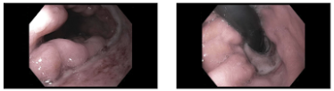
Methods: A 68-year-old female with a history of breast cancer undergoing active chemotherapy (last cycle 10 days prior), schizophrenia, and chronic cannabis use presented with worsening altered mental status and recurrent nausea and vomiting. She had multiple recent admissions for similar symptoms with only partial relief from proton pump inhibitors (PPIs) and sucralfate. Laboratory findings included leukocytosis (WBC 25.3 K/uL), iron-deficiency anemia (Hb dropped from 7.4 to 6.9 g/dL, requiring transfusion), thrombocytosis (Plt 661 K/uL), and elevated liver enzymes (ALT 71 U/L, ALP 193 U/L). CT chest revealed lower esophageal wall thickening with paraesophageal fluid, raising suspicion for esophagitis or malignancy. Esophagogastroduodenoscopy (EGD) revealed LA Grade D esophagitis, an oozing esophageal ulcer, and a distinct polypoid mucosal mass in the lower esophagus. Biopsies of the mass showed ulcerated granulation tissue with acute inflammation and were positive for CMV via immunostaining. The patient was treated with oral valganciclovir, alongside continued PPI and sucralfate therapy. Discussion: This case highlights an atypical presentation of CMV esophagitis as an esophageal mass, contrasting with its more common ulcerative manifestations. In immunocompromised patients presenting with esophageal masses, CMV infection should be considered in the differential diagnosis to avoid misdiagnosis with malignancy or other conditions. Prompt and accurate diagnosis is crucial, as antiviral therapy is effective. This case underscores the importance of thorough histopathological evaluation, including CMV immunostaining, in such clinical scenarios.
Figure: Polypoid Mucosal Mass In the Lower Esophagus
Disclosures: Seyma Bayram indicated no relevant financial relationships. Mutaz Kalas indicated no relevant financial relationships. Mehmet Talha Bayram indicated no relevant financial relationships. Amit Sohagia indicated no relevant financial relationships.
Seyma Bayram, MD1, Mutaz Kalas, MD2, Mehmet Talha Bayram, MD3, Amit Sohagia, MD4. P0679 - CMV Esophagitis Presenting as an Esophageal Mass in an Immunocompromised Patient, ACG 2025 Annual Scientific Meeting Abstracts. Phoenix, AZ: American College of Gastroenterology.