
Sunday Poster Session
Category: Colon
 photo")
Muhammad Abdullah, MBBS (he/him/his)
Lahore Medical and Dental College
Talagang, Punjab, Pakistan
Lymphoglandular complexes (LGCs) in the colonic submucosa are characterized by epithelial glands embedded within the submucosal lymphoid aggregates. Adenomatous polyps may rarely extend into these complexes, resulting in a pseudo-invasion where dysplastic glands appear in the submucosa. It may pose a significant diagnostic challenge as this histologic pattern closely mimics invasive adenocarcinoma. We present a case of a dysplastic rectal polyp with pseudo-invasion of submucosal LGCs simulating superficial invasive adenocarcinoma.
Case Description/
Methods:
A 61-year-old male underwent a screening colonoscopy, where a single rectal lesion with ulceration was identified and biopsied, with pathology revealing focal active colitis with low-grade dysplasia. A follow-up diagnostic colonoscopy showed similar findings, prompting further evaluation. A CT scan of the abdomen and pelvis showed no evidence of metastatic disease or lymphadenopathy. The patient subsequently underwent flexible sigmoidoscopy with endoscopic submucosal dissection (ESD). A 22 mm polypoidal lesion was identified in the rectum, 12 cm proximal to the anal verge. The lesion was superficial and elevated, with some central depression, indicating a Paris classification IIa. Histology revealed high-grade dysplasia and intramucosal adenocarcinoma with glandular elements extending into submucosal LGC, initially raising concern for superficial invasion. However, further review by multiple pathologists favored pseudo-invasion into pre-existing LGCs. Immunohistochemistry demonstrated intact mismatch repair protein expression. The patient recovered uneventfully and was encouraged to follow up in 6 months for a surveillance colonoscopy.
Discussion:
The involvement of LGCs by colonic adenomatous dysplasia presents a rare phenomenon that is most commonly observed in the rectum due to the higher density of LGCs. It is pertinent to distinguish benign colon polyps from true invasive adenocarcinoma through histopathological cues, including the absence of desmoplasia and lamina propria preservation. In our case, flexible sigmoidoscopy with ESD successfully resected the lesion en bloc, following which an expert review confirmed glandular pseudo-invasion. This case illustrates the importance of careful histopathological evaluation of benign mimics such as pseudo-invasion into LGCs to avoid unnecessary invasive intervention.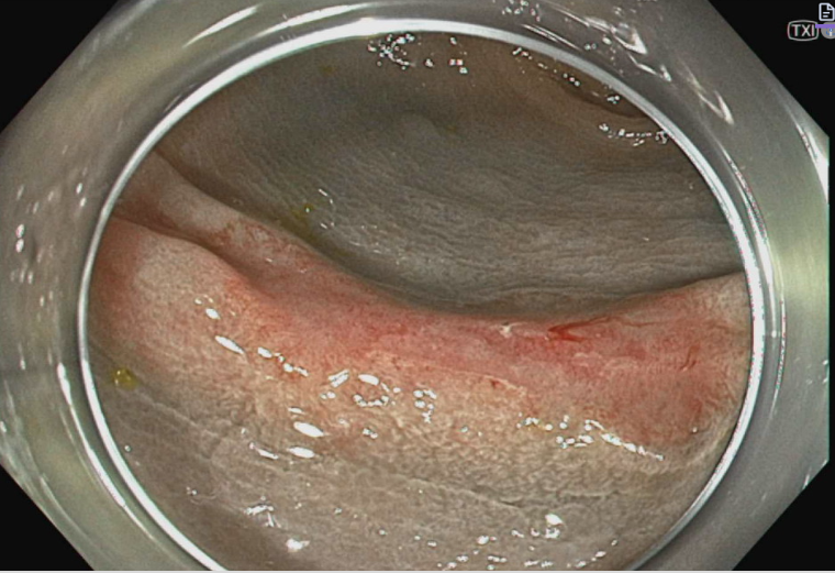
Figure: Figure 1. Endoscopic view of the flexible sigmoidoscopy showing a 22 mm polypoidal, ulcerated lesion in the rectum, 12 cm proximal to the anal verge.
Disclosures:
Muhammad Abdullah indicated no relevant financial relationships.
Abdullah Javed indicated no relevant financial relationships.
Muhammad Aftab indicated no relevant financial relationships.
Charanjeet Singh indicated no relevant financial relationships.
Muhammad Abdullah, MBBS1, Abdullah Javed, MBBS2, Muhammad Aftab, MD3, Charanjeet Singh, MD4. P0432 - The Great Pretender: A Case of Pseudoinvasion of Lymphoglandular Complexes by a Dysplastic Rectal Adenoma, ACG 2025 Annual Scientific Meeting Abstracts. Phoenix, AZ: American College of Gastroenterology.