
Sunday Poster Session
Category: Colon
 photo")
Shreya Deshpande (she/her/hers)
SS Institute of Medical Sciences and Research Center, Davangere
Bangalore, Karnataka, India
We report a case of bullous pemphigoid (BP) in a 74-year-old male diagnosed with colon adenocarcinoma, where the temporal relationship between the BP and cancer suggests an association between the two, though controversial.
Case Description/
Methods:
A 74-year-old male with a history of a cerebrovascular accident 20 years ago, requiring hemicraniectomy presented with worsening diffuse skin blisters and desquamation. Dermatology was consulted and punch biopsies from the left arm confirmed BP. He was treated initially with intravenous steroids and was transitioned to oral steroids gradually. During hospitalization, his hemoglobin dropped to 6.1 g/dL from his baseline of 11 g/dL, and 4 units of packed red blood cells were transfused. He had an episode of hematochezia, and gastroenterology was consulted. Colonoscopy demonstrated a circumferential, nearly obstructing mass thought to be in the transverse colon and biopsies were taken. CT imaging revealed bilateral renal lesions that were concerning for renal cell carcinoma, but no other sites concerning for malignancy. Carcinoembryonic antigen levels were normal. Pathology revealed moderately differentiated adenocarcinoma without microsatellite instability. Colorectal surgery was consulted, and surgical resection of the malignancy was planned.
Discussion:
BP is a polymorphic autoimmune blistering skin condition commonly affecting the elderly. Known triggers include immune dysregulation, infections, trauma and neurologic diseases. The association of BP and the presence of underlying malignancies is controversial. However, there have been studies showing a higher incidence of malignancy in patients with BP, making this a compelling topic. In our case, the diagnosis of colon adenocarcinoma followed a recent diagnosis of an uncommon dermatosis without a clear inciting factor. The discovery of underlying malignancy could have resulted in immune dysregulation, potentially acting as a trigger for BP. We present an interesting case of concomitant development of BP and colon cancer for which an association warrants further study.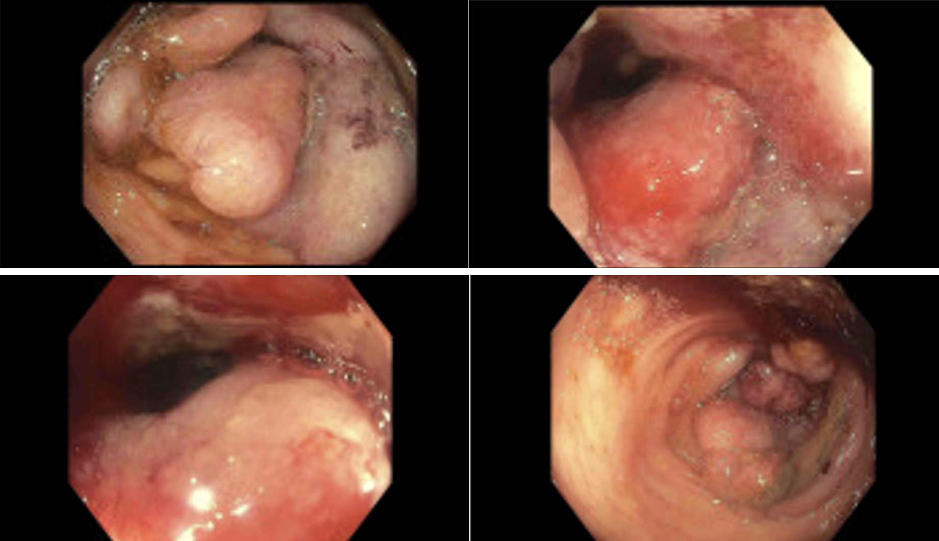
Figure: Figure 1. Representative colonoscopy images of mass in the transverse colon nearly obstructing the lumen. 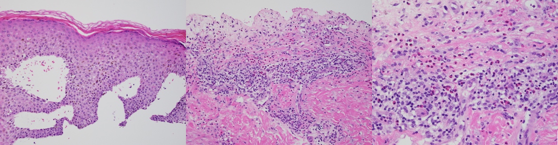
Figure: Figure 2. Dermatopathology of Bullous Pemphigoid.
Left panel. SEBulla 400X magnification: Epidermis showing a subepidermal bulla with complete separation from the underlying dermis. Center panel. Eos 200X magnification: Superficial dermal inflammatory infiltrate with numerous eosinophils. Right panel. Eos 400X magnification: Dermal inflammatory infiltrate with numerous eosinophils
Disclosures:
Shreya Deshpande indicated no relevant financial relationships.
Akshay Chandora indicated no relevant financial relationships.
Aaron Hein indicated no relevant financial relationships.
Elnaz Jafarimehr indicated no relevant financial relationships.
Shreya Deshpande, 1, Akshay Chandora, MD2, Aaron Hein, MD3, Elnaz Jafarimehr, MD3. P0423 - Blistering Surprises: A Rare Association Between Bullous Pemphigoid and Colon Adenocarcinoma, ACG 2025 Annual Scientific Meeting Abstracts. Phoenix, AZ: American College of Gastroenterology.