University of North Dakota, School of Medicine and Health Sciences Fargo, ND
Award: ACG Presidential Poster Award
Guneet Sidhu, MD1, Emilee Ohman, BS1, Hussam Almasri, MD2, Alexey Hodkoff, MD3, John Bassett, MD3 1University of North Dakota, School of Medicine and Health Sciences, Fargo, ND; 2University of North Dakota, School of Medicine and Health Sciences, West Fargo, ND; 3Sanford Health, Fargo, ND Introduction: Specimen provenance complications, including tissue contamination and misidentification, represent a critical challenge in surgical pathology with potential for significant clinical and ethical consequences. Despite strict protocols, cross-contamination can occur at various stages of specimen handlingduring grossing, embedding, or slide preparationleading to inaccurate diagnoses. Short tandem repeat (STR) DNA analysis has emerged as a gold standard for resolving such discrepancies by providing definitive tissue identity verification
Case Description/
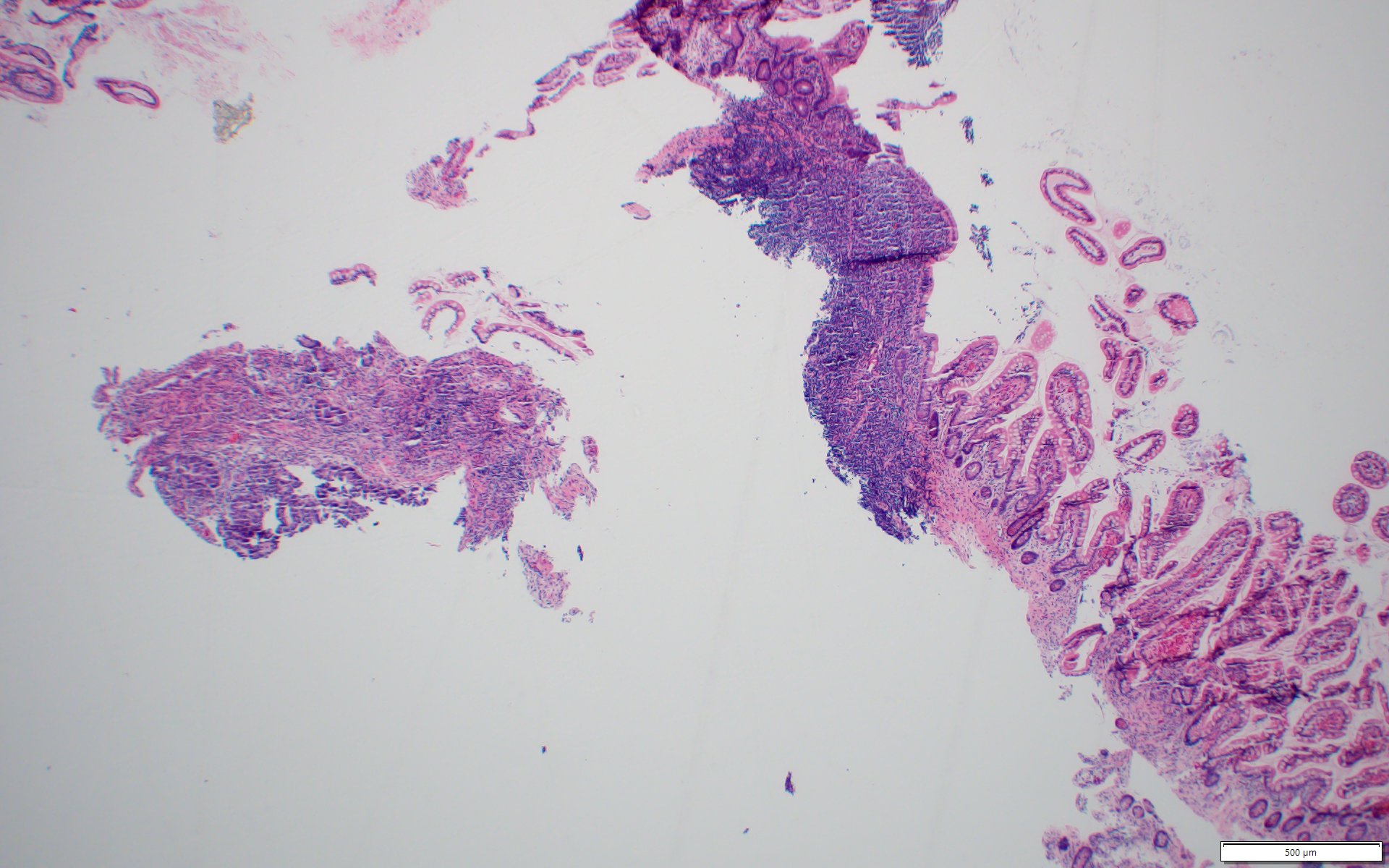
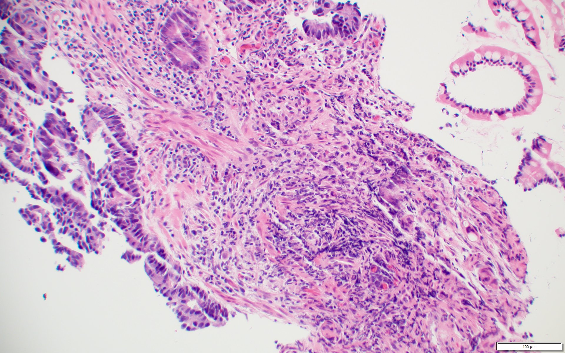
Methods: A 31-year-old male with a history of psoriasis, allergic rhinitis, and indeterminate colitis diagnosed in 2019 presented for routine surveillance colonoscopy. His initial diagnosis followed a 3-year history of fecal urgency and frequency, with endoscopic findings consistent with ulcerative proctosigmoiditis. He was started on mesalamine, leading to sustained symptomatic remission. A follow-up colonoscopy in January 2025 showed no evidence of inflammation, mass, or mucosal abnormalities. However, histopathologic evaluation of a terminal ileum biopsy unexpectedly revealed a small fragment of tissue with adenocarcinoma adjacent to unremarkable ileal mucosa. Fig. A shows a low magnificationunremarkable ileal mucosa on the right and a fragment of adenocarcinoma on the left. Fig B higher magnification image of infiltrative cell nests with intracytoplasmic mucin in the lower right corner. Given the complete lack of endoscopic or clinical evidence of malignancy, the sample underwent DNA fingerprinting using STR polymorphism analysis, which confirmed that the adenocarcinoma tissue originated from a different patient. The contamination was most likely introduced at the grossing station or during tissue processing in the laboratory.
Discussion: This case underscores the vital role of molecular tissue identity testing in modern pathology. STR analysis has proven highly effective in identifying specimen contamination and resolving tissue provenance errors, with wide applications in both clinical and forensic settings. In this instance, it prevented a potentially devastating misdiagnosis that could have led to patient distress, unnecessary repeat colonoscopy, or even unwarranted oncologic intervention. Pathologists and clinicians must maintain a high index of suspicion when pathological findings are inconsistent with clinical or endoscopic impressions, and STR testing should be considered in such scenarios to ensure diagnostic accuracy
Figure: Low magnification image with an unremarkable ileal mucosa on the right and a fragment of tissue with adenocarcinoma on the left
Figure: Higher magnification image of the tissue fragment with adenocarcinoma, including infiltrative cell nests with intracytoplasmic mucin in the right lower corner
Disclosures: Guneet Sidhu indicated no relevant financial relationships. Emilee Ohman indicated no relevant financial relationships. Hussam Almasri indicated no relevant financial relationships. Alexey Hodkoff indicated no relevant financial relationships. John Bassett indicated no relevant financial relationships.
Guneet Sidhu, MD1, Emilee Ohman, BS1, Hussam Almasri, MD2, Alexey Hodkoff, MD3, John Bassett, MD3. P0415 - When Biopsy Lies: DNA Fingerprinting Exposes Hidden Contaminant, ACG 2025 Annual Scientific Meeting Abstracts. Phoenix, AZ: American College of Gastroenterology.