
Sunday Poster Session
Category: Colon
 photo")
Ashley Serjilus, MD (she/her/hers)
Naval Medical Center Portsmouth
Portsmouth, VA
Gastrointestinal (GI) subepithelial lesions (SEL) are due to tissue growth arising underneath the intestinal mucosa. They are identified on 1 in 300 endoscopies, often incidentally, and range from benign to malignant etiologies. Heterotopic pancreas (HP) an uncommon developmental anomaly where pancreatic tissue occurs outside of the pancreas organ. We present a case of an incidentally found SEL in the jejunum of an otherwise healthy young female found to be HP on surgical resection.
Case Description/
Methods:
A 25-year-old woman presented to the emergency department with recurrent right upper quadrant (RUQ) abdominal pain and nausea, particularly after fatty meals. A computed tomography (CT) scan confirmed cholelithiasis, and she underwent a laparoscopic cholecystectomy, which resolved her RUQ pain. The CT also incidentally revealed a 15mm hyperattenuating mass in the jejunum. Further testing, including a Dotatate position emission tomography (PET) scan did not identify any avid lesions. Laboratory testing of chromogranin A and carcinoembryonic antigen (CEA) levels were normal. Push enteroscopy identified a 15 mm submucosal lesion in the jejunum. Biopsies of the overlying mucosa were normal. Interventional Gastroenterology was referred for consideration of an Endoscopic Ultrasound (EUS), but due to the distal nature of the lesion, it was not recommended. The patient underwent surgical resection of the lesion which revealed heterotopic pancreatic tissue without malignancy.
Discussion:
Further evaluation of incidental findings of SELs on endoscopy to determine benign versus malignant etiology is imperative. While often lesions can be evaluated through EUS, the location within the GI tract may limit EUS as a diagnostic tool and surgical resection or biopsy is required. While most SELs are asymptomatic, depending on location and size, they can cause pain, GI bleeding or obstruction. Diagnosis of HP is challenging as imaging alone cannot be used to confirm the findings. In this case, surgical resection revealed HP as the underlying etiology of an incidental SEL. While also often asymptomatic, there are cases of HP causing pancreatitis, dyspepsia, or GI bleeding. With an incidence of 0.11-0.21% in autopsies, over 90% of cases are reported to involve the stomach, duodenum, or jejunum. Despite this, jejunal SELs from HP are reported rarely in the literature. This case adds to the literature of how a jejunal SEL due to HP can appear radiographically and endoscopically in an asymptomatic patient.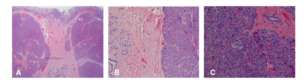
Figure: A: 20x – Pancreatic acini and ducts below small bowel mucosa.
B: 100x – Closely packed pancreatic acini next to an area of ductules and ducts
C: 200x – Pancreatic acinar cells with a ductule.
Disclosures:
Ashley Serjilus indicated no relevant financial relationships.
Christopher Thorndal indicated no relevant financial relationships.
Ashley Matias indicated no relevant financial relationships.
Mercy Wagner indicated no relevant financial relationships.
Rebecca Johnson indicated no relevant financial relationships.
Allison Bush indicated no relevant financial relationships.
Ashley Serjilus, MD1, Christopher Thorndal, DO1, Ashley Matias, MD1, Mercy Wagner, MD1, Rebecca Johnson, MD1, Allison Bush, MD2. P0400 - Subepithelial Surprise: A Rare Care of Heterotopic Pancreatic Tissue in the Jejunum, ACG 2025 Annual Scientific Meeting Abstracts. Phoenix, AZ: American College of Gastroenterology.