Diana V. Vera-Garcia, MD1, Yingyun Yang, MD1, Paul Travers, MD1, Michael B. Wallace, MD1, Dilhana Badurdeen, MBBS, MD2 1Mayo Clinic, Jacksonville, FL; 2Mayo Clinic, Jacksoville, FL Introduction: Ewing's sarcoma typically presents in the bones of children and adolescents. Extra-skeletal cases, particularly in the perineum, are exceedingly rare, with only a couple reported worldwide. Due to its rarity, the diagnosis poses a significant challenge, requiring a high degree of clinical suspicion. Here, we describe a unique case of Ewing's sarcoma originating in the perineum of an adult patient.
Case Description/
Methods: A 36-year-old male presented with a 2-year history of rectal bleeding. He experienced rectal pain and blood after wiping that was relieved by showering without other symptoms. The patient's past medical history and laboratory tests were unremarkable. He denied experiencing weight change, anorexia, fatigue, or night sweats. His family history was negative for relevant gastrointestinal conditions. Pelvic MRI reported a transsphincteric fistula from the lower anal canal, coursing to the left medial peritoneum (~4 cm). Additionally, a 2.1 x 1.2 cm T2 hyperintense, avidly enhancing lesion was observed within the subcutaneous fat of the right perineum. Colonoscopy confirmed a left-sided perianal fistula in the intergluteal area, without drainage. An ultrasound-guided soft tissue biopsy showed an oval hypoechoic lesion in the subcutaneous fat of the right perineum. The obtained pathology samples were positive for soft tissue myoepithelioma, with nuclear atypia but no mitotic activity. Subsequent immunohistochemistry staining demonstrated positive expression of keratin and S100, indicative of a myoepithelial neoplasm with S100 positivity and EWSR1 translocation. This diagnosis was further validated by FISH analysis, which detected a rearrangement involving the EWSR1 gene region, consistent with Ewing's Sarcoma. Anorectal examination under anesthesia confirmed a left-sided anterior fistula. The tract was surgically debrided, and a silastic seton was placed. Given the pathology findings of a slowly growing tumor, the patient was referred for perineal mass excision and to mental health services for additional care. Discussion: Although exceptionally rare, perineal Ewing's sarcoma should be considered as a differential diagnosis of soft tissue tumors arising in the perineum. This case exemplifies the complexities associated with this disease, diagnosis, and management. A coordinated approach involving healthcare professionals is essential to improve patient outcomes and ensure holistic care.
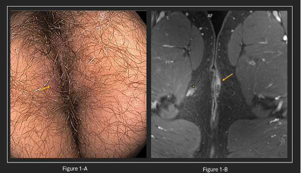
Figure: Figure 1-A: Colonoscopy showing a perianal fistula on the left side. Figure 1-B: Axial MRI with an arrow indicating a transsphincteric fistula; the "x" marks the enhancing lesion on the right perineum.
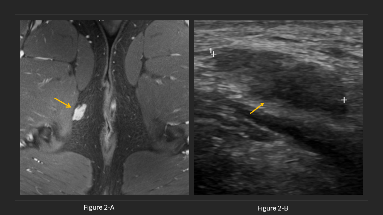
Figure: Figure 2-A: shows a T2 hyperintense enhancing lesion within the subcutaneous fat of the right perineum. Figure 2-B: depicts the same lesion observed through ultrasound.
Disclosures: Diana Vera-Garcia indicated no relevant financial relationships. Yingyun Yang indicated no relevant financial relationships. Paul Travers indicated no relevant financial relationships. Michael Wallace indicated no relevant financial relationships. Dilhana Badurdeen indicated no relevant financial relationships.
Diana V. Vera-Garcia, MD1, Yingyun Yang, MD1, Paul Travers, MD1, Michael B. Wallace, MD1, Dilhana Badurdeen, MBBS, MD2. P0399 - Extra Skeletal Ewing’s Sarcoma Where You Least Expect It: A Perineal Manifestation, ACG 2025 Annual Scientific Meeting Abstracts. Phoenix, AZ: American College of Gastroenterology.

 photo")