Ingrid Rocha, BS1, Suraj Suresh, MD2 1Wayne State School of Medicine, Detroit, MI; 2Henry Ford Health, Detroit, MI Introduction: Colonic extranodal marginal zone lymphoma of mucosa-associated lymphoid tissue (MALT lymphoma, ML) is a rare and diagnostically challenging entity with variable morphology. Although molecular analysis can aid diagnosis, it is often discovered incidentally, and both its pathogenesis and optimal treatment remain poorly defined. We present a case of ML identified within a diminutive colonic polyp during evaluation for liver transplant in a patient with cryptogenic cirrhosis.
Case Description/
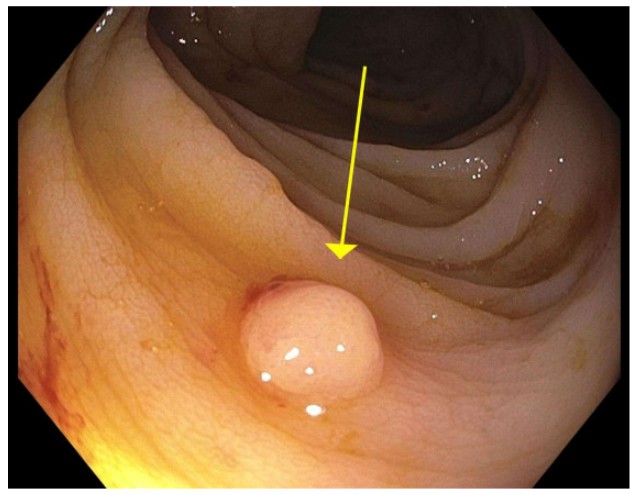
Methods: A 62-year-old male with biopsy-proven cryptogenic cirrhosis, rare Pi-FZ alpha-1 antitrypsin deficiency, and heterozygous HFE C282Y mutation was admitted for worsening anemia. He reported hematochezia and epistaxis the day prior to admission and denied anticoagulant use. An EGD two months prior showed portal hypertensive gastropathy. A colonoscopy three years earlier revealed sigmoid diverticulosis and a non-dysplastic polyp. On admission, he was stable with an unremarkable exam. Labs showed hemoglobin 8.2 g/dL and platelets 90 K/μL. Repeat EGD showed mild gastropathy, and gastric biopsies were negative for H. pylori. Colonoscopy revealed friable mucosa, external hemorrhoids, and a 4 mm transverse colon polyp (Figure 1). Histologic analysisof the resected polyp demonstrated CD5/10-negative low-grade B-cell lymphoma with plasmacytic differentiation and clonal B-cell gene rearrangement, consistent with MALT lymphoma. Bone marrow biopsy showed trilineage hematopoiesis and low-level gelatinous transformation. PET imaging was denied by insurance. Due to the absence of a safe window for lymph node biopsy, the patient is currently undergoing surveillance with serial imaging to monitor for disease progression before reactivation on the liver transplant list. Discussion: To our knowledge, this represents the first reported case of colonic MALT lymphoma in a patient with cryptogenic cirrhosis. Colonic ML is a rare and often incidental finding, frequently diagnosed only through removal and histologic evaluation of diminutive polyps. This case highlights the diagnostic challenges of localizing and characterizing such lesions and raises the possibility of a previously unrecognized association between cirrhosis and colonic ML. Further investigation into the underlying pathophysiology of colonic MALT lymphoma and its potential link to liver disease is warranted.
Figure: Figure 1: 4 mm polyp resected from the transverse colon.
Disclosures: Ingrid Rocha indicated no relevant financial relationships. Suraj Suresh indicated no relevant financial relationships.
Ingrid Rocha, BS1, Suraj Suresh, MD2. P0394 - Incidental Colonic Polyp Uncovers MALT Lymphoma During Transplant Work-Up, ACG 2025 Annual Scientific Meeting Abstracts. Phoenix, AZ: American College of Gastroenterology.

 photo")