
Sunday Poster Session
Category: Colon
Faisal Mehmood, MD
HonorHealth
Glendale, AZ
The association between right-sided colon cancer and acute appendicitis was first reported by Shears in 1906. The incidence of acute appendicitis as an initial sign of cecal or ascending colon cancer ranges from 3.4% to 15%. When both conditions coexist, symptoms of appendicitis tend to be more prominent. We present a case of a young male who underwent appendectomy and was later found to have metastatic colon cancer.
Case Description/
Methods:
A 34-year-old male presented with stabbing right lower quadrant pain, similar to his episode of acute appendicitis three months prior. He was febrile to 100.4°F, tachycardic at 119 bpm, and hypertensive to 161/113 mmHg. Labs revealed leukocytosis (19,000/µL) and mild acute kidney injury with creatinine 1.5 mg/dL. Liver enzymes, electrolytes, and coagulation panel were within normal limits. Computed Tomography (CT) abdomen & pelvis with IV contrast revealed a new thickening of the ascending colon and distal ileum, and multifocal low-density liver lesions (Figure 1).
Liver abscesses were initially suspected. An ultrasound-guided biopsy of the liver lesions was performed, revealing metastatic adenocarcinoma with widespread necrosis, consistent with colonic origin (Figure 2). Immunohistochemical staining showed positivity for AE1/AE3, CK20, CDX2, and SATB2, indicating a colorectal origin of the malignancy (Figure 3). He had no family history of colorectal cancer and had never undergone prior endoscopic evaluation. Colonoscopy revealed a large, ulcerated, partially obstructing fungating mass in the cecum (Figure 4). Biopsies confirmed poorly differentiated invasive adenocarcinoma (Figure 5). Immunohistochemistry for mismatch repair proteins demonstrated intact nuclear expression of MLH1, MSH2, MSH6, and PMS2. A diverting colostomy was performed laparoscopically, and he was discharged with close oncology follow-up.
Discussion:
Colorectal cancer (CRC) after appendectomy is increasingly reported, not only in patients over 40 but also among younger adults, as illustrated by this case. CRC often develops within six months post-appendectomy. While studies debate the utility of postoperative colonoscopy in those over 40, no clear guidelines exist. With the rising incidence of CRC in younger populations, acute appendicitis is being considered a possible early warning sign. This supports ongoing discussion about whether all patients aged 18–59 with acute appendicitis should undergo colonoscopy, as CT imaging alone may miss early malignancy.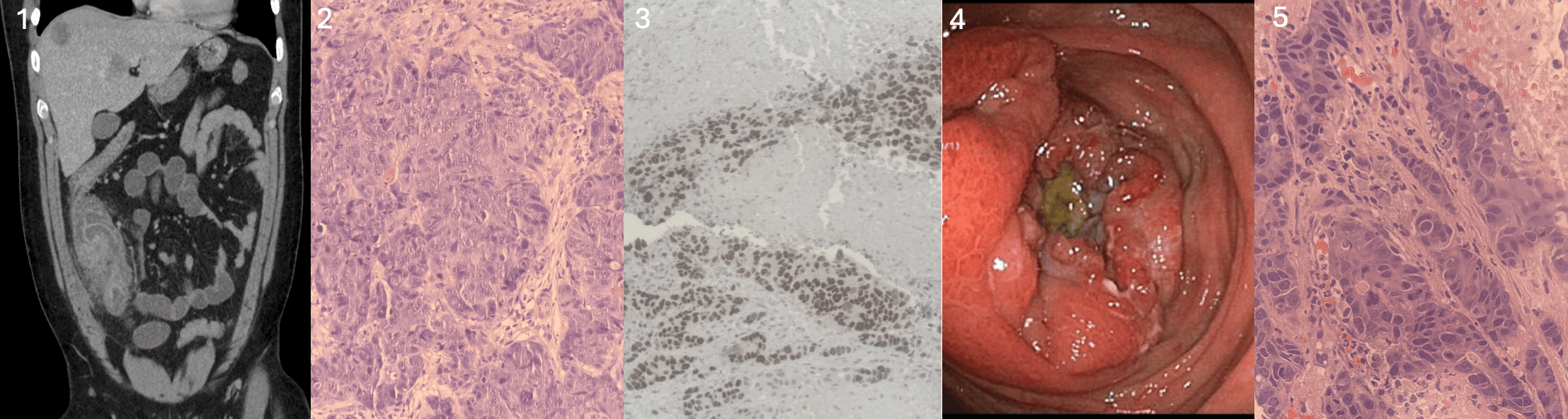
Figure: Figure 1). CT abdomen & pelvis with IV contrast showing a new thickening of the ascending colon and distal ileum, and multifocal low-density liver lesions.
Figure 2). Liver biopsy showing infiltrative gland-forming malignant neoplasm characterized by atypical architecture and nuclear atypia with frequent mitotic figures and extensive tumoral necrosis.
Figure 3). Immunohistochemical stain is SATB2, showing strong nuclear positivity, suggestive of colorectal origin of malignancy.
Figure 4). Colonoscopy showing a large, ulcerated, partially obstructing fungating mass in the cecum.
Figure 5). Colon mass biopsy showing infiltrative gland-forming malignant neoplasm characterized by cytologically malignant cells with increased nuclear to cytoplasmic ratios and architectural disarray.
Disclosures:
Hajra Jamil indicated no relevant financial relationships.
Faisal Mehmood indicated no relevant financial relationships.
Mehul Patel indicated no relevant financial relationships.
Gavin Levinthal indicated no relevant financial relationships.
Hajra Jamil, MD1, Faisal Mehmood, MD2, Mehul Patel, DO3, Gavin Levinthal, MD4. P0392 - Metastatic Colon Cancer Following Appendectomy Mimicking Liver Abscess, ACG 2025 Annual Scientific Meeting Abstracts. Phoenix, AZ: American College of Gastroenterology.