
Sunday Poster Session
Category: Colon
Faisal Mehmood, MD
HonorHealth
Glendale, AZ
Mucosal neuromas are rare, benign neural lesions located within the colonic mucosa. They are typically asymptomatic and most often discovered incidentally during a routine colonoscopy. Here, we present a case of intramucosal neuroma in a middle-aged man who underwent a screening colonoscopy.
Case Description/
Methods:
A 61-year-old male presented for colorectal cancer screening at an outpatient surgery center. His past medical history included hypertension, obesity, and an anxiety disorder. Physical examination was unremarkable. Laboratory tests, including a complete blood count and comprehensive metabolic panel, were within normal limits. His previous colonoscopy, performed at age 50, revealed no polyps.
During the current procedure, colonoscopy revealed a 4 mm semi-sessile polyp in the sigmoid colon, which was removed using a cold snare technique, with complete resection and retrieval (Figure 1). Histopathological analysis showed colonic mucosa with a subtle intramucosal spindle cell proliferation, characterized by bland spindled cells with elongated, wavy nuclei, consistent with an intramucosal neuroma (Figure 2). Immunohistochemical staining demonstrated strong nuclear positivity for S100, and negative staining for CD117 (C-Kit) (Figure 3).
Discussion:
Benign nerve sheath tumors are most commonly found in the skin and soft tissues. Their presence in the gastrointestinal tract is exceedingly rare. In the colon, these lesions may present as small polyps or mass-like formations. The differential diagnosis includes mucosal neuromas (including those associated with Multiple Endocrine Neoplasia type 2B [MEN-2B]), neurofibromas (characteristic for neurofibromatosis type 1 [NF-1]), schwannomas, ganglioneuromas (linked to NF-1 and Cowden syndrome), and gastrointestinal stromal tumors (GISTs).
It is important to distinguish mucosal neuromas from these entities based on immunohistochemical markers. Neuromas are positive for S100 and negative for C-Kit (CD117) and DOG-1, markers that are typically expressed in GISTs. Treatment consists of local excision, which is curative. There is no specific surveillance protocol for mucosal neuromas; however, standard colonoscopy surveillance guidelines should be followed based on the type, number, and histological features of polyps identified.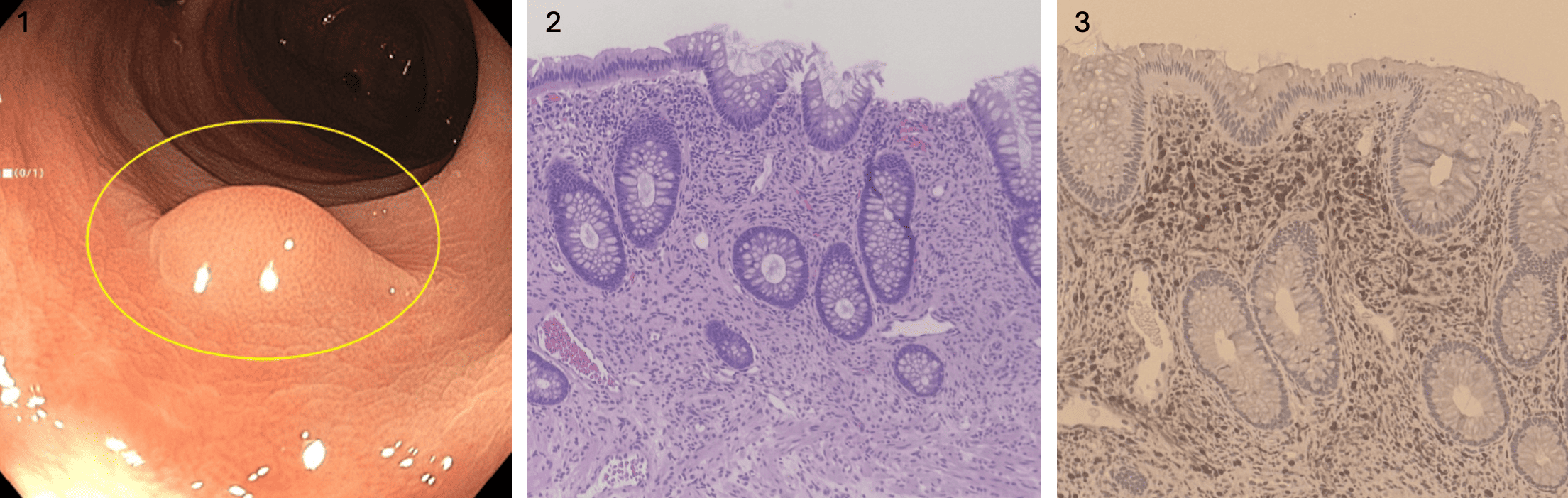
Figure: Figure 1: A 4 mm semi-sessile polyp in the sigmoid colon.
Figure 2: Histopathological analysis of the polyp showed colonic mucosa with a subtle intramucosal spindle cell proliferation, characterized by bland spindled cells with elongated, wavy nuclei, consistent with an intramucosal neuroma.
Figure 3: Immunohistochemical staining demonstrated strong nuclear positivity for S100.
Disclosures:
Hajra Jamil indicated no relevant financial relationships.
Faisal Mehmood indicated no relevant financial relationships.
Joseph Fares indicated no relevant financial relationships.
Hajra Jamil, MD1, Faisal Mehmood, MD2, Joseph Fares, MD3. P0391 - Intramucosal Neuroma: A Rare Neural Lesion Discovered During Routine Colonoscopy, ACG 2025 Annual Scientific Meeting Abstracts. Phoenix, AZ: American College of Gastroenterology.