
Sunday Poster Session
Category: Colon
Rehan Rafiq, MD, FACG (he/him/his)
Mercy Hospital Jefferson
Festus, MO
A 48-year-old African American man presented for routine index colonoscopy. Past medical history was significant for marginal zone lymphoma in remission and metastatic (Stage T4 N1). CDRCC treated with left nephrectomy and chemotherapy. He was asymptomatic. Social and family history was unremarkable. Prior to colonoscopy, a CT of the abdomen and pelvis showed no metastatic disease. The colonoscopy revealed a 5 cm descending colon mass causing partial luminal obstruction. Biopsy of colon mass showed CDRCC. Patient underwent left hemicolectomy. On one year follow up, the patient is asymptomatic, and imaging demonstrated no evidence of recurrent CDRCC.
Discussion:
Renal cell cancers typically present in the 6th and 7th decades of life, with male to female ratio of 2:115. 4% of these tumors are familial16. CDRCC is a rare variant of CCRCC3. The CDRCC is more common in African Americans and presents with locally advanced (T3-4) disease, distant metastasis and nodal involvement3. 3-year disease specific survival for CDRCC is 58%3,17.
CDRCC metastasizes to lymph nodes, lungs, pleura, bones and liver2,18. Colon metastasis of CDRCC in our case, is a novel finding. Thin slice CT has higher sensitivity for detecting local recurrence and metastatic disease5,19,20. CT scan findings of medullary location, weak and heterogeneous enhancement, infiltrative growth, preserved renal contour and cystic component may predict ductal cell histology21,22. Histologically, CDRCC exhibits high grade tubular morphology and infiltrative growth accompanied by stromal desmoplasia2. Surgery is the mainstay of the treatment. Treatment for metastatic disease is chemotherapy2. 5 years survival after nephrectomy is poor23. The median survival is 16 months2.
This case underscores the rarity of metastatic CDRCC to the colon. Since no previous case of CDRCC with metastasis to colon has been documented, this finding may be prognostic.
Figure: From left to right: descending colon mass, hemotoxylin and eosin (H&E) stain of biopsied colon mass showing stromal desmoplasia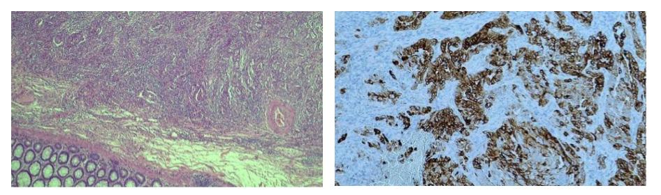
Figure: From left to right: H&E stain of tumor and normal colon, immunohistochemical stain (IHC) of tumor
Disclosures:
Rehan Rafiq indicated no relevant financial relationships.
Umar Rafiq indicated no relevant financial relationships.
Nancy Muller indicated no relevant financial relationships.
Muhammad Yousaf indicated no relevant financial relationships.
Rehan Rafiq, MD, FACG1, Umar Rafiq, 2, Nancy Muller, MD3, Muhammad Yousaf, MD4. P0383 - Colonic Metastasis of Renal Collecting Duct Carcinoma: An Unusual Presentation, ACG 2025 Annual Scientific Meeting Abstracts. Phoenix, AZ: American College of Gastroenterology.