Muhammad Arsalan, MD1, Wagmah Javed. Khan, MD1, Mustafa Shah, MD2, Hameed Ullah, MD3, Jahangir Khan. Afridi, MD4, Elie Chahla, MD1 1St. Luke's Hospital, Chesterfield, MO; 2Geisinger Health System, Danville, PA; 3St Luke’s Hospital, Chesterfield, Chesterfield, MO; 4St. Luke's Hospital Missouri, Chesterfield, MO Introduction: Mucosa associated lymphoid tissue (MALT) lymphoma is most commonly found in the stomach and is often associated with H. pylori infections. Manifestations outside the stomach are very rare and have an unclear etiology. In the colon, if rarely present, it usually presents as a polyp. We present a case of a primary colonic MALT lymphoma (< 0.5% of all colon cancers) presenting as benign-appearing colonic submucosal nodules, an appearance distinct from the commonly observed polypoid appearance.
Case Description/
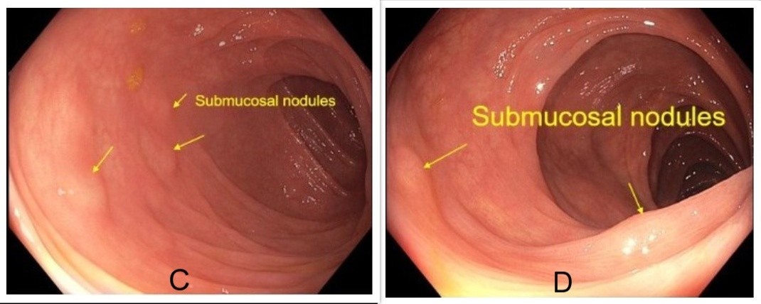
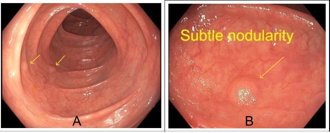
Methods: A 63-year-old asymptomatic man with normal routine blood work underwent a screening colonoscopy, which revealed multiple subtle submucosal nodules in the sigmoid colon, ascending colon, transverse colon and cecum. Biopsies and histopathological analysis of the ascending colon nodules revealed low-grade B-cell non-Hodgkin lymphoma, consistent with extra nodal marginal zone MALT lymphoma, further supported by immunohistochemical staining. PET imaging revealed thickening of the cecum and proximal ascending colon, concerning for lymphoma infiltration, along with enlarged right mesocolic, central mesenteric and retroperitoneal lymph nodes. Bone marrow biopsy revealed minimal involvement. Based on these findings, the patient was staged as Stage IV extra nodal marginal zone lymphoma (MZL). Treatment was initiated with rituximab and lenalidomide and continued for over a year. A follow-up colonoscopy almost 2 years later showed complete resolution of the submucosal nodules, with random biopsies confirming the absence of residual or recurrent disease. Discussion: Primary colonic MALT lymphoma is a rare variant, comprising only 2.5% of all MALT lymphomas. The cecum and ascending colon are the most involved sites, with up to 70% of cases occurring proximally to the hepatic flexure. Usually, it is asymptomatic but can present with GI bleeding or abdominal pain. Colonoscopy may reveal polypoid or benign-looking nodular lesions, posing a risk of being overlooked during routine screening due to its subtle nodular presentation. This case highlights the asymptomatic nature and nonspecific presentation of MALT lymphomas. It emphasizes the importance of keeping a low threshold for obtaining biopsies of even benign-appearing submucosal nodules in the lining of the colon, as early detection of colonic MALT lymphoma can allow curative treatment and lead to more favorable patient outcomes.
Figure: Image A: Submucosal nodules in the transverse colon. Image B: Subtle submucosal nodules in the ascending colon.
Figure: Image C: Submucosal nodules in the sigmoid colon. Image D: Submucosal nodules in the ascending colon.
Disclosures: Muhammad Arsalan indicated no relevant financial relationships. Wagmah Khan indicated no relevant financial relationships. Mustafa Shah indicated no relevant financial relationships. Hameed Ullah indicated no relevant financial relationships. Jahangir Afridi indicated no relevant financial relationships. Elie Chahla indicated no relevant financial relationships.
Muhammad Arsalan, MD1, Wagmah Javed. Khan, MD1, Mustafa Shah, MD2, Hameed Ullah, MD3, Jahangir Khan. Afridi, MD4, Elie Chahla, MD1. P0346 - Primary Colonic MALT Lymphoma Presenting as Submucosal Nodules: A Rare Presentation, ACG 2025 Annual Scientific Meeting Abstracts. Phoenix, AZ: American College of Gastroenterology.