Ronan Allencherril, MD, Mary R. Schwartz, MD, Thomas R. McCarty, MD, MPH Houston Methodist Hospital, Houston, TX Introduction: Strongyloides stercoralis is an intestinal parasite found globally that infrequently presents with disseminated disease in immunocompetent patients. Infections are often confined to the intestinal lumen, with rare involvement of the biliary tract. We present a case of biliary strongyloidiasis presenting with ampullary thickening on imaging.
Case Description/
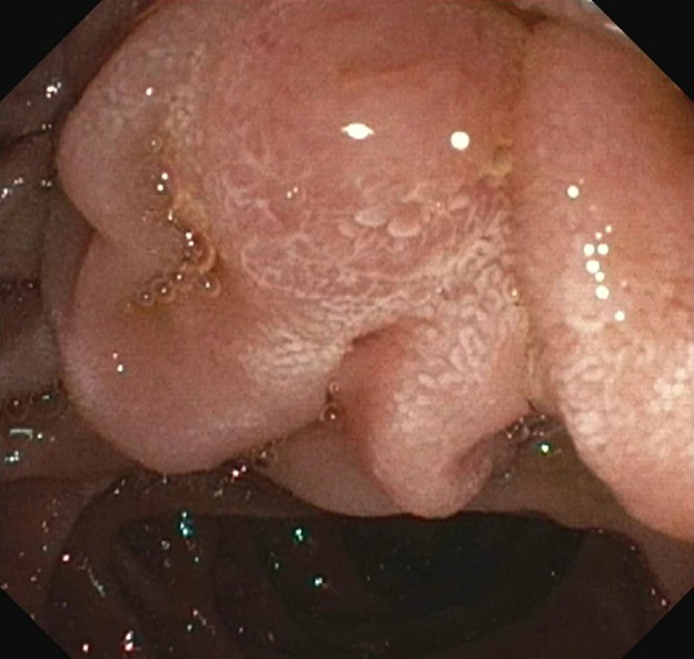
Methods: An 84-year-old gentleman with a history of obstructive sleep apnea, cholelithiasis status-post cholecystectomy, and pancreas cysts presented due to an abnormal magnetic resonance cholangiopancreatography (MRCP). He reported chronic epigastric pain refractory to proton pump inhibitor (PPI) and a 70 lb weight loss over the past 2 years. He was hospitalized for epigastric pain and found to have worsening intrahepatic biliary dilation and a possible lesion of the ampulla. Labs showed normal liver chemistries and a peripheral eosinophilia. MRCP showed a double duct sign with ampullary thickening and stable pancreatic head cysts. The decision was made to proceed with endoscopic ultrasound (EUS) and endoscopic retrograde cholangiopancreatography (ERCP). Upper endoscopy showed congested periampullary mucosa (Figure 1). EUS revealed atrophy and hyperechoic strands of the pancreas with mildly dilated pancreatic duct and a 20 mm septated cystic lesion in the pancreatic head which was sampled by fine needle aspiration (FNA). On EUS, the common bile duct was 12 mm with mild narrowing at the lower third of the common bile duct with an abrupt tapering at the distal main duct. There was wall thickening of the ampulla on EUS. On ERCP, the lower third of the main bile duct contained a mild stenosis with upstream dilation. Sphincterotomy and balloon sweep were performed with removal of sludge. Brushings were sent for cytology and one plastic stent was placed in the common bile duct. Cytology returned with larvae compatible with S. stercoralis and the patient was treated with ivermectin (Figure 2). Stool ova and parasite testing also showed S.stercoralis. FNA cytology confirmed intraductal papillary mucinous neoplasm and the biliary stent was removed on subsequent ERCP. After treatment with ivermectin, his abdominal pain resolved and he was discharged with no further concerns. Discussion: This case shows a unique presentation of biliary Strongyloides in an immunocompetent patient without environmental risk factors. The case enforces the importance of sampling indeterminate biliary strictures as well as differential for peripheral eosinophilia.
Figure: Edematous ampulla on EGD
Figure: Cytology via brushings demonstrating Strongyloides stercoralis
Disclosures: Ronan Allencherril indicated no relevant financial relationships. Mary Schwartz indicated no relevant financial relationships. Thomas McCarty: Covidien/Medtronic – Consultant. Endoquest Robotics – Consultant.
Ronan Allencherril, MD, Mary R. Schwartz, MD, Thomas R. McCarty, MD, MPH. P0153 - Biliary Strongyloidiasis Presenting With an Ampullary Lesion on MRI, ACG 2025 Annual Scientific Meeting Abstracts. Phoenix, AZ: American College of Gastroenterology.