Sunday Poster Session
Category: Biliary/Pancreas
P0110 - Spontaneous Choledochoduodenal Fistula Post-Cholecystectomy: Successful Endoscopic Management of a Rare Complication Using Cholangioscopy and Lithotripsy
Sunday, October 26, 2025
3:30 PM - 7:00 PM PDT
Location: Exhibit Hall

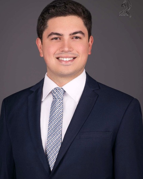
Fernando Lugo-Hernandez, MD (he/him/his)
HCA Florida Healthcare
Brandon, FL
Presenting Author(s)
Fernando Lugo-Hernandez, MD1, Omar Jureyda, DO2, Viraj Modi, DO1, Bhavtosh Dedania, MD1
1HCA Florida Healthcare, Brandon, FL; 2HCA Florida Healthcare, Tampa, FL
Introduction: Spontaneous choledochoduodenal fistula (CDF) is a rare complication of chronic choledocholithiasis, particularly in post-cholecystectomy patients. Management is often challenging due to anatomic distortion, stone burden, and increased procedural complexity. We present a unique case of post-cholecystectomy cholangitis with incidental spontaneous CDF and refractory bile duct stones, successfully managed with advanced endoscopic techniques including direct visualization cholangioscopy and electrohydraulic lithotripsy (EHL).
Case Description/
Methods: 75-year-old woman with hypertension, diabetes, and prior cholecystectomy presented with confusion, lethargy, fever, epigastric pain, and vomiting. Labs showed leukocytosis (15.5 × 10⁹/L), transaminitis (AST 780, ALT 458 U/L), total bilirubin 2.2 mg/dL, and lactate 2.8 mmol/L. CT and ultrasound revealed bile duct dilation and choledocholithiasis. She met Tokyo Guidelines 2018 criteria for grade II cholangitis.
Initial ERCP revealed a spontaneous choledochoduodenal fistula, periampullary diverticulum, and multiple CBD stones. A biliary sphincterotomy, balloon sweep, and placement of one plastic and one bare metal stent were performed. Stones were seen exiting through the fistula, bypassing the ampulla. She improved but was readmitted two weeks later with recurrent cholangitis and persistent ductal stones on imaging.
A second ERCP with direct visualization cholangioscopy revealed large, densely calcified stones. EHL was used for fragmentation, but multiple balloon catheters ruptured due to stone burden. A distal CBD stricture was noted. A fully covered self-expanding metal stent was placed for both biliary drainage and fistula tamponade. The patient improved, with normalization of liver enzymes and resolution of symptoms.
Discussion: This case highlights a rare instance of spontaneous CDF in a post-cholecystectomy patient. Fistula formation likely stemmed from prolonged biliary obstruction and chronic inflammation. Endoscopic management was complicated by altered anatomy, periampullary diverticulum, and a high stone burden. Cholangioscopy enabled direct visualization and successful EHL. Covered metal stenting maintained ductal patency and facilitated fistula closure. This approach avoided the need for surgery in a high-risk elderly patient.
In select patients, minimally invasive, advanced endoscopic modalities such as cholangioscopy and EHL can provide definitive therapy, even in complex biliary anatomy, and may obviate surgical intervention.
