Sneha Rao Adidam, MD, Lakshmi Chirumamilla, MD, Joseph Asemota, MD, Amro Abdellatief, MD, Justice Arhinful, MD, Angesom Kibreab, MD, Farshad Aduli, MD Howard University Hospital, Washington, DC Introduction: Hemosuccus pancreaticus occurs due to fistulous communication between the pancreatic duct and peripancreatic vessel. It has a mortality of about 9.6%. Diagnosis may be challenging; given its rarity, 1 in every 1500 cases of GI bleeding, and obscure presentation with intermittent bleeding. Herein, the authors report cases that illustrate the diagnostic challenge in 2 middle aged patients.
Case Description/
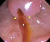
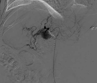
Methods: Case 1: A 69-year-old lady with history of alcohol use disorder and pancreatitis presented with abdominal pain and hematemesis with a Hb of 5.3g/dL. 2 units PRBCs were transfused and EGD showed esophagitis and gastritis, but no evidence of active or recent bleeding. Hospital course was complicated by hematemesis and hematochezia. Repeat endoscopy showed blood in the second portion of duodenum without any active source of bleeding. Hence, a side viewing endoscope was used, and intermittent bleeding was noted from the ampulla of Vater (see Image 1). CTA was immediately done and revealed pseudoaneurysm of the common hepatic artery, which was embolized (see Image 2). She remained clinically stable with no further bleeding and was discharged home.
Case 2: A 55-year-old male with medical history of alcohol use disorder, chronic pancreatitis, and vocal cord squamous cell cancer presented with 2 weeks of abdominal pain, melena and a hemoglobin of 5.4g/dL. He was transfused and CT chest/abdomen/pelvis showed a 6 x 4 x 5 cm mass adjacent to the tail the pancreas with enhancing central mass representing a splenic artery or pancreatic artery pseudoaneurysm. EGD showed no active bleeding and prominent ampulla of Vater. Despite embolization of the splenic artery pseudoaneurysm by IR, patient continued to have bloody stools. Repeat EGD showed extrinsic mass with ulcerated mucosa without any active bleeding. Immediate angiography revealed bleeding from splenic artery pseudoaneurysm and left gastric artery. Angioembolization was performed with no further bleeding. Discussion: Hemosuccus pancreaticus is extremely rare and requires high index of suspicion. Endoscopic visualization of active bleeding is only evident in 30% of cases but the diagnostic yield can be increased with a side viewing endoscope. Angiography remains the gold standard with a sensitivity of >96% for diagnostic and therapeutic embolization of bleeding aneurysms.
Figure: Image 1. Side viewing endoscope with evidence of blood from the ampulla of Vater
Figure: Image 2. Angioembolization of the common hepatic artery
Disclosures: Sneha Rao Adidam indicated no relevant financial relationships. Lakshmi Chirumamilla indicated no relevant financial relationships. Joseph Asemota indicated no relevant financial relationships. Amro Abdellatief indicated no relevant financial relationships. Justice Arhinful indicated no relevant financial relationships. Angesom Kibreab indicated no relevant financial relationships. Farshad Aduli indicated no relevant financial relationships.
Sneha Rao Adidam, MD, Lakshmi Chirumamilla, MD, Joseph Asemota, MD, Amro Abdellatief, MD, Justice Arhinful, MD, Angesom Kibreab, MD, Farshad Aduli, MD. P5286 - A Case Series of Hemosuccus Pancreaticus: The Silent Bleeders, ACG 2025 Annual Scientific Meeting Abstracts. Phoenix, AZ: American College of Gastroenterology.

 photo")