P0212 - Multifocal Solid Pseudopapillary Neoplasm in an Adolescent Female With a Positive Family History of Familial Adenomatous Polyposis: A Rare Case Report
King Faisal Specialist Hospital and Research Centre Riyadh, Ar Riyad, Saudi Arabia
Abdulrahman Alharbi, MBBS1, Hend Almuhaya, MBBS1, Bader Alajlan, 1, Aymen Almuhaidb, MD2 1King Faisal Specialist Hospital and Research Centre, Riyadh, Ar Riyad, Saudi Arabia; 2King Faisal Specialist Hospital and Research Centre, Ar Riyad, Ar Riyad, Saudi Arabia Introduction: The coexistence of familial adenomatous polyposis (FAP) and pancreatic solid pseudopapillary neoplasms (SPN) is extremely uncommon, with limited reports in the literature. SPN is a low-grade malignant tumor, usually solitary and seen in young females. Multifocal SPN is exceedingly rare, and its occurrence in the setting of FAP background has not been well described.
Case Description/
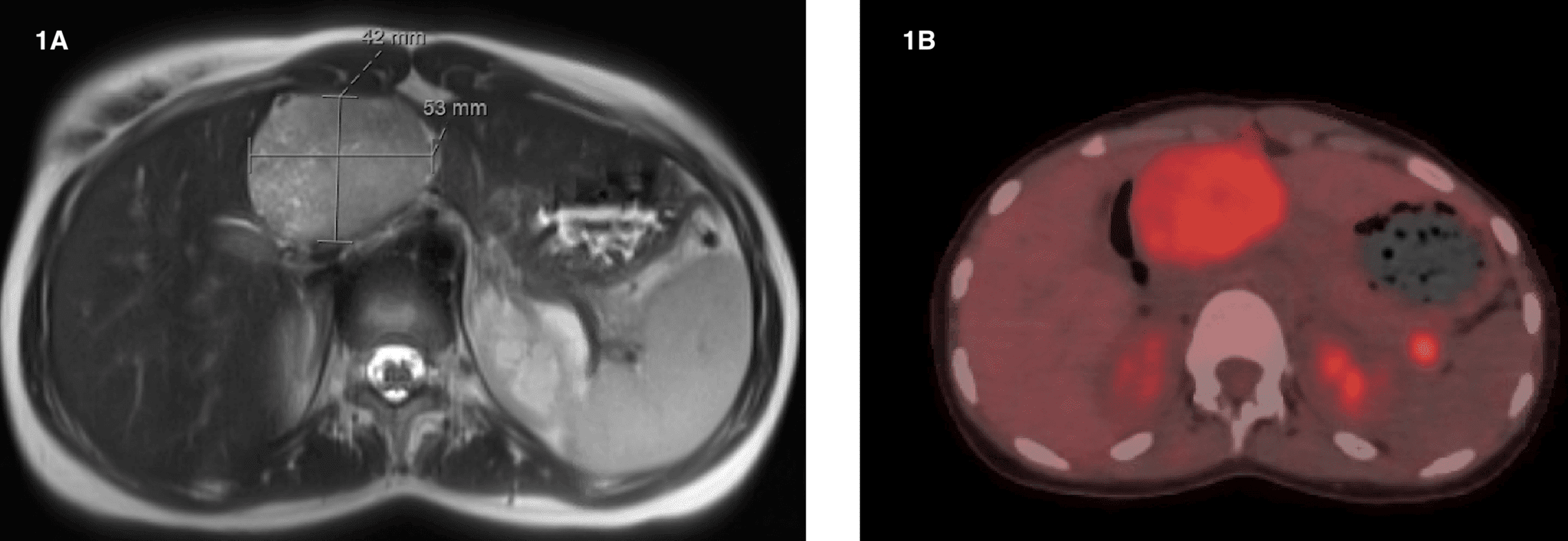
Methods: A 15-year-old female diagnosed with FAP was referred to our hospital for further evaluation. The patient reported two months of intermittent epigastric pain, nausea, and unintentional weight loss of 2 kilograms. Magnetic resonance imaging (MRI) abdomen revealed multiple enhancing pancreatic lesions, raising suspicion for multifocal SPN (Figure 1A). Contrast-enhanced computed tomography (CT) abdomen, showed 4 well-defined, hypoenhancing intrapancreatic lesions ranging from 0.7 to 1.3 cm and a 5.7 cm hypovascular mass in the porta hepatis compressing the portal confluence and associated with collateral formation. A positron emission tomography (PET) scan was performed (Figure 1B). Esophagogastroduodenoscopy was normal. Endoscopic ultrasound (EUS) identified a large heterogeneous, hypervascular, hypoechoic, multicystic mass in the porta hepatis. EUS-guided fine needle biopsy (FNB) was performed. Cytology immunostaining revealed nuclear β-catenin positivity, loss of E-cadherin membranous expression, CD99 exhibited dot-like positivity, and progesterone receptor (PR) was positive while synaptophysin showed weak positivity, findings consistent with SPN. Following a multidisciplinary tumor board discussion, the patient underwent open central pancreatectomy with Roux-en-Y pancreaticojejunostomy and cholecystectomy. Histopathology confirmed multifocal SPN, staged as pT3 N0, with negative margins and no lymphovascular or nodal involvement. The gallbladder and excised lymph node were benign. Recovery was uneventful. Genetic testing and colonoscopy for FAP surveillance are pending. Discussion: This case illustrates an unusual presentation of multifocal SPN in an adolescent female with a background of familial cancer predisposition. While SPNs are generally considered sporadic, the possible association with FAP, though rare, warrants further investigation. The case also highlights the importance of a high index of suspicion, advanced imaging modalities, and early multidisciplinary collaboration in managing rare pancreatic tumors. Awareness of such rare presentations can aid early recognition.
Figure: Figure 1A: MRI abdomen showed multiple pancreatic lesions, largest seen at the pancreatic head measures 5.3 x 4.2 cm abutting first part of duodenum and compressing portal vein. No biliary dilatation or hepatic lesions. Figure 1B: PET scan demonstrated mild fluorodeoxyglucose (FDG) uptake in the pancreatic masses and indeterminate metabolic activity in the sigmoid colon
Disclosures: Abdulrahman Alharbi indicated no relevant financial relationships. Hend Almuhaya indicated no relevant financial relationships. Bader Alajlan indicated no relevant financial relationships. Aymen Almuhaidb indicated no relevant financial relationships.
Abdulrahman Alharbi, MBBS1, Hend Almuhaya, MBBS1, Bader Alajlan, 1, Aymen Almuhaidb, MD2. P0212 - Multifocal Solid Pseudopapillary Neoplasm in an Adolescent Female With a Positive Family History of Familial Adenomatous Polyposis: A Rare Case Report, ACG 2025 Annual Scientific Meeting Abstracts. Phoenix, AZ: American College of Gastroenterology.