University of South Florida Morsani College of Medicine Tampa, FL
Rishi Bolla, MD1, Kevin Shah, MD1, Susan Jacob, MD1, Edward O'Neill, MD2, Samuel Slone, MD1 1University of South Florida Morsani College of Medicine, Tampa, FL; 2Tampa General Hospital, Tampa, FL Introduction: Mucosal Schwann cell hamartomas (MSCH) are benign tumors characterized by Schwann cell hyperproliferation in the lamina propria of the colon. They are exceedingly rare and are distinct from other peripheral nerve sheath tumors due to their lack of associated syndromic conditions such as neurofibromatosis (NF) or multiple endocrine neoplasia (MEN) syndrome. They are generally asymptomatic and often identified incidentally during screening colonoscopy, however recognition is crucial to differentiate them from other lesions of the gastrointestinal tract to avoid unnecessary interventions.
Case Description/
Methods: A 72-year-old male had a colonoscopy three years prior during which eight tubular adenomatous polyps were resected. He presented fora follow up surveillance colonoscopy which was performedwithoutcomplications. He was found to have six polyps throughout the colon which were resected. Histologic examination of a polyp found in the descending colon demonstrated spindle cell proliferation in the lamina propria; immunohistochemistry was positive for S100 and SOX-10 and negative for EMA, GLUT-1, CD34, DOG-1, and desmin. These findings were consistent with MSCH. The remainder of the polyps found were tubular adenomas. Discussion: Our patient does not have history of NF or MEN syndrome and denied any family history of conditions associated with neural lesions. It is unclear whether there is an increased chance of developing MSCH if other adenomatous polyps are present in the colon. Furthermore, the rarity of MSCH precludes any meaningful study, and examination of the few cases reported in literature failed to reveal any genetic or environmental risk factors. Since MSCH is a benign lesion, it is important that it is recognized by clinicians in order to save the patient from unnecessary workup or intervention.
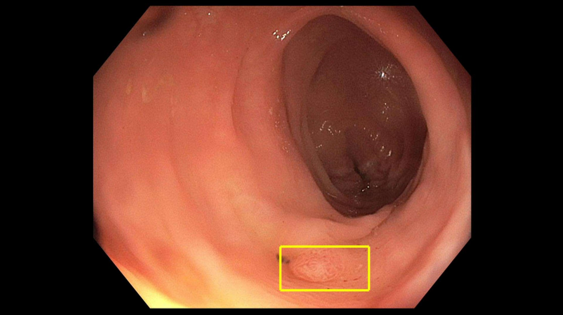
Figure: Figure 1: Polyp found in the descending colon.
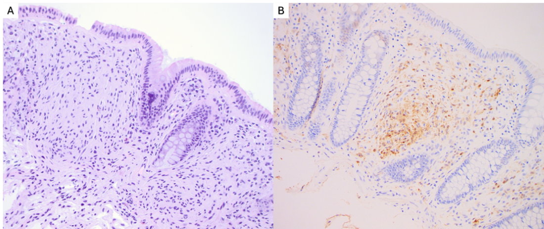
Figure: Figure 2: A) H&E slide showing morphology (20x magnification). B) S100 immunohistochemical stain, which is positive in the lesion cells (20x magnification).
Disclosures: Rishi Bolla indicated no relevant financial relationships. Kevin Shah indicated no relevant financial relationships. Susan Jacob indicated no relevant financial relationships. Edward O'Neill indicated no relevant financial relationships. Samuel Slone indicated no relevant financial relationships.
Rishi Bolla, MD1, Kevin Shah, MD1, Susan Jacob, MD1, Edward O'Neill, MD2, Samuel Slone, MD1. P0376 - A Rare Case of Mucosal Schwann Cell Hamartoma Found in the Descending Colon, ACG 2025 Annual Scientific Meeting Abstracts. Phoenix, AZ: American College of Gastroenterology.