Georgetown University School of Medicine Washington, D.C., DC
Ahmad Abdulraheem, MD1, Neha Tallapragada, BA2, Walid Chalhoub, MD3, Salwan Almashat, MD4, Omar Abureesh, MD5, Usman Afzal, MD4 1MedStar Washington Hospital Center-Georgetown University, Washington, DC; 2Georgetown University School of Medicine, Washington, D.C., DC; 3MedStar Georgetown University Hospital, Washington, DC; 4MedStar Health, Washington, D.C., DC; 5Staten Island University Hospital, Northwell Health, Staten Island, NY Introduction: Ganglioneuromas (GNs) are benign neuroblastic tumors derived from neural crest cells or mature ganglion cells, often involving the sympathetic ganglia. Common sites include the posterior mediastinum, adrenal gland, and retroperitoneum. Gastrointestinal (GI) GNs are rare, and most reported cases involve diffuse colonic involvement related to familial conditions like MEN2B or NF1. Isolated ascending colonic GNs are uncommon, with fewer than 15 reported cases managed by polypectomy alone.We report a sporadic ascending colon GN polyp treated with endoscopic resection.
Case Description/
Methods: A 48-year-old asymptomatic male underwent routine screening colonoscopy. He had no personal or family history of colorectal cancer or polyposis syndromes and denied tobacco, alcohol, or recreational drug use. Colonoscopy revealed a 5 mm sessile, pinkish polyp in the ascending colon. Histopathological analysis of the initial biopsy identified a GN.
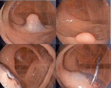
A repeat colonoscopy was performed two months later for complete resection. Due to the sessile morphology and need for definitive histological assessment, hot snare polypectomy via endoscopic mucosal resection (EMR) was performed (Figure 1). Gross examination revealed a dome-shaped, tan-pink lesion measuring 1.1 × 0.8 × 0.8 cm.
Immunohistochemical staining was positive for S100 and synaptophysin, confirming the diagnosis of GN. Final pathology demonstrated a mucosal polypoid ganglioneuroma with hyperplastic changes (Figure 2). The patient was advised to undergo surveillance colonoscopy in one year. Discussion: GNs are rare, slow-growing benign neoplasms that are typically discovered incidentally during endoscopic procedures. They are often identified in pathology by confirming ganglion cell involvement via immunostains such as chromogranin A, neurofilament, S100, and/or synaptophysin. In sporadic, localized cases such as the one presented, endoscopic resection followed by routine surveillance is typically sufficient for management. In contrast, diffuse or syndromic cases may require more invasive management and genetic counseling.
Figure: Figure 1: Colonoscopy images demonstrate a 1 cm sessile polyp in the ascending colon (top panels). Post-resection images show the site after hot snare endoscopic mucosal resection (EMR) with clip placement (bottom panels).