Wright Center for Graduate Medical Education Scranton, PA
Sunny Kumar, MD1, Satish Kumar Ahuja, MD1, Jyoti Yadav, MD2, Anila Mahesh, MD3, Danial Nadeem, MD4, Nihaal Karnik, MD4, Joseph Bannon, MD5, Lucas Murray, MD4, Zongxian Cao, MD6 1Wright Center for Graduate Medical Education, Scranton, PA; 2The Wright Center for Graduate Medical Education, Scranton, PA; 3Geisinger Wyoming Valley Medical Center, Duryea, PA; 4Geisinger Wyoming Valley Medical Center, Wilkes-Barre, PA; 5Geisinger Community Medical Center, Scranton, PA; 6Geisinger Wyoming Valley Medical Center, Danville, PA Introduction: Small cell neuroendocrine carcinoma (SCNEC) of the gastrointestinal tract is a rare and highly aggressive malignancy characterized by rapid progression and early metastasis. Unlike the more common adenocarcinomas of the colon and rectum, neuroendocrine tumors may present with atypical symptoms and can be diagnostically challenging due to their heterogeneous pathology. Here, we describe the case of a middle-aged female whose initial nonspecific presentation belied an advanced and aggressive malignancy, ultimately confirmed as SCNEC.
Case Description/
Methods: A 45-year-old female with no significant past medical history presented with a 3-week history of constipation and rectal bleeding. She denied weight loss, urinary symptoms, or a family history of cancer. Labs, (CBC) Complete Blood Count showed thrombocytosis (platelets 449 K/µL). CT (Computed Tomography) abdomen revealed an 8.1 cm pelvic mass abutting the rectum, along with hepatic lesions, peritoneal nodules, a sclerotic iliac lesion, and a left hydrosalpinx, suggestive of metastatic disease. MRI (Magnetic Resonance Imaging) pelvis demonstrated a circumferential rectal mass invading mesorectal fat and adjacent lymphadenopathy.
Colonoscopy revealed an ulcerated mass from 6 to 12.5 cm from the anal verge. Initial biopsies showed only mucosal fragments with focal atypia. PET (Positron Emission Tomography) scan confirmed hypermetabolic activity in the pelvis, bones, upper abdomen, and adnexa. Flexible sigmoidoscopy with deeper biopsy was performed, which confirmed the diagnosis of SCNEC. The patient was started on systemic etoposide and cisplatin. She was deemed ineligible for surgery due to extensive metastases, and palliative radiation was considered. Discussion: This case highlights the aggressive nature of rectal SCNEC and the importance of high clinical suspicion when faced with atypical GI presentations. The National Comprehensive Cancer Network (NCCN) and the European Neuroendocrine Tumor Society (ENETS) support platinum-based chemotherapy as first-line treatment for metastatic high-grade NECs, as was initiated in this case. Surgery is reserved for localized disease; hence, it is not pursued here. Prognosis for metastatic SCNEC remains poor, with a median survival of 10–16 months. Early diagnosis and accurate histopathology are critical to initiating appropriate therapy and optimizing outcomes.
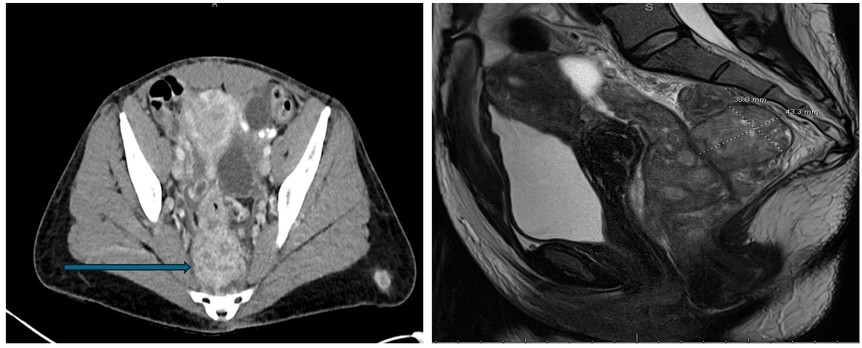
Figure: Image 1A: CT showing an 8.1 cm pelvic mass abutting the right rectum. Image 1B: MRI showing a partially circumferential rectal tumor measuring 5.4 cm in length.
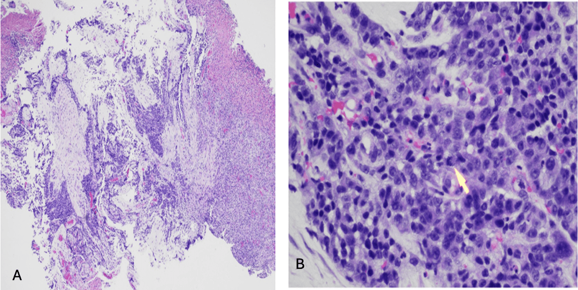
Figure: Image 2A: Lower magnification showed ulcerated rectal mucosa involved by infiltrated tumor composed of strands and sheets of small epithelioid cells. Image 2B: Higher magnification showed tumor cells with scant cytoplasm, irregular nuclear contours, inconspicuous nucleoli, and frequent mitotic activity [two mitotic figures in the center, one highlighted with an arrow].
Disclosures: Sunny Kumar indicated no relevant financial relationships. Satish Kumar Ahuja indicated no relevant financial relationships. Jyoti Yadav indicated no relevant financial relationships. Anila Mahesh indicated no relevant financial relationships. Danial Nadeem indicated no relevant financial relationships. Nihaal Karnik indicated no relevant financial relationships. Joseph Bannon indicated no relevant financial relationships. Lucas Murray indicated no relevant financial relationships. Zongxian Cao indicated no relevant financial relationships.
Sunny Kumar, MD1, Satish Kumar Ahuja, MD1, Jyoti Yadav, MD2, Anila Mahesh, MD3, Danial Nadeem, MD4, Nihaal Karnik, MD4, Joseph Bannon, MD5, Lucas Murray, MD4, Zongxian Cao, MD6. P0531 - A Rare Case of Metastatic Small Cell Neuroendocrine Carcinoma of the Rectum, ACG 2025 Annual Scientific Meeting Abstracts. Phoenix, AZ: American College of Gastroenterology.

 photo")