University of Miami Miller School of Medicine at Holy Cross Hospital Fort Lauderdale, FL
John Coleman, MD1, Alana J. Kosches, BA2, Ingrid C.. Landfald, MD3, Paul Feldman, MD4, Daniel S.. Kosches, MD5 1University of Miami Miller School of Medicine at Holy Cross Hospital, Fort Lauderdale, FL; 2SUNY Downstate Health Sciences University, Brooklyn, NY; 3Department Of Clinical Anatomy Mazovian Academy, Płock, Poland, Poland, Mazowieckie, Poland; 4Holy Cross Medical Group, Plantation, FL; 5Department of Gastroenterology, University of Miami Miller School of Medicine, Holy Cross Health Hospital, Fort Lauderdale, FL Introduction: Dieulafoy lesions are a rare but significant cause of upper gastrointestinal (GI) bleeding, accounting for approximately 6% of cases. These lesions are often challenging to diagnose, especially when the initial esophagogastroduodenoscopy (EGD) is non-diagnostic.
Case Description/
Methods: We present the case of a 77-year-old female with a history of cardiovascular and gastrointestinal diseases who presented with acute blood loss anemia and hypotension. Initial EGD was non-diagnostic, but a subsequent computerized tomography with angiography (CTA) revealed an active bleed from a Dieulafoy lesion. This finding guided a successful repeat endoscopy with hemostatic clipping, leading to stabilization of the patient. Discussion: This case highlights the importance of CTA in conjunction with EGD for diagnosing and managing elusive Dieulafoy lesions, particularly in high-risk patients. The multimodal approach outlined in this case adds to existing literature by demonstrating how imaging can complement standard endoscopic management, ultimately improving diagnostic accuracy and patient outcomes.
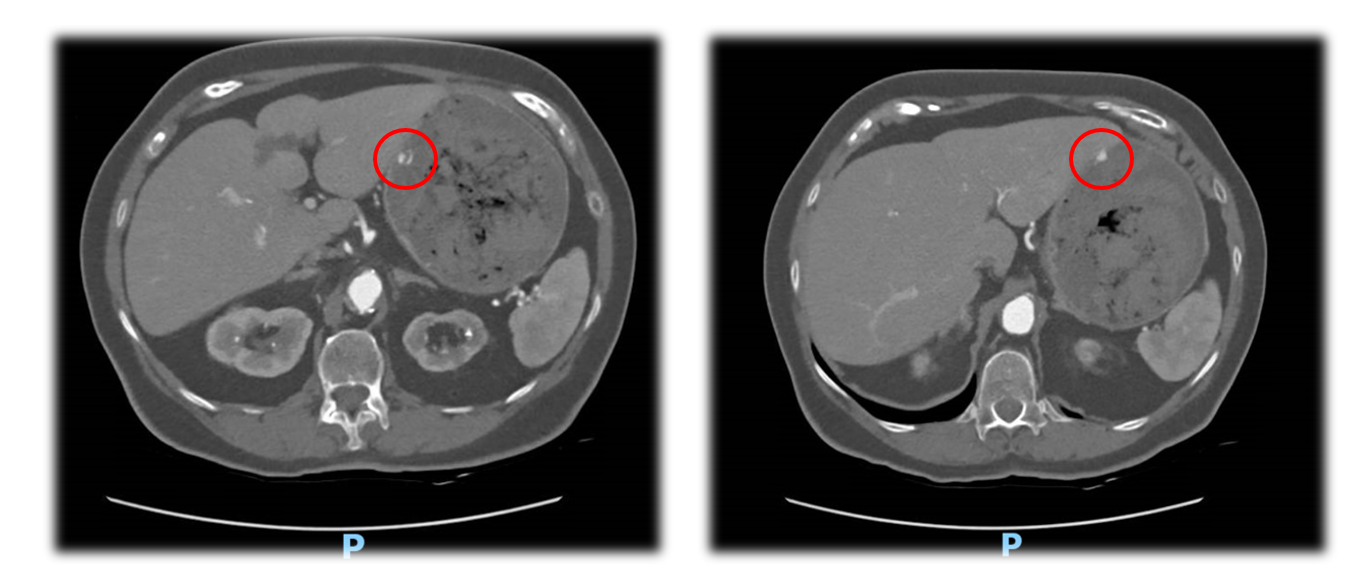
Figure: Figure 1a and 1b: Axial view images at two different cuts from an abdominal computed tomography angiography scan on initial patient presentation revealing a small actively oozing Dieulafoy lesion (red circle, left and right) in the proximal anterior segment of the stomach.
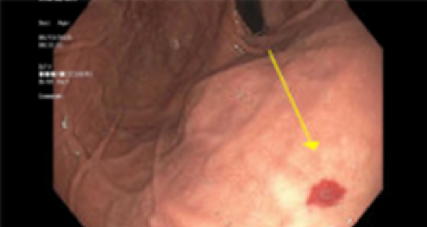
Figure: Figure 2: Captured image of Dieulafoy lesion (yellow arrow, above) during second EGD with active oozing in the proximal anterior segment of the stomach.
Disclosures: John Coleman indicated no relevant financial relationships. Alana Kosches indicated no relevant financial relationships. Ingrid Landfald indicated no relevant financial relationships. Paul Feldman indicated no relevant financial relationships. Daniel Kosches indicated no relevant financial relationships.
John Coleman, MD1, Alana J. Kosches, BA2, Ingrid C.. Landfald, MD3, Paul Feldman, MD4, Daniel S.. Kosches, MD5. P0970 - Active Gastrointestinal Bleeding From Dieulafoy Lesion on Abdominal Computed Tomography Angiography Imaging, ACG 2025 Annual Scientific Meeting Abstracts. Phoenix, AZ: American College of Gastroenterology.