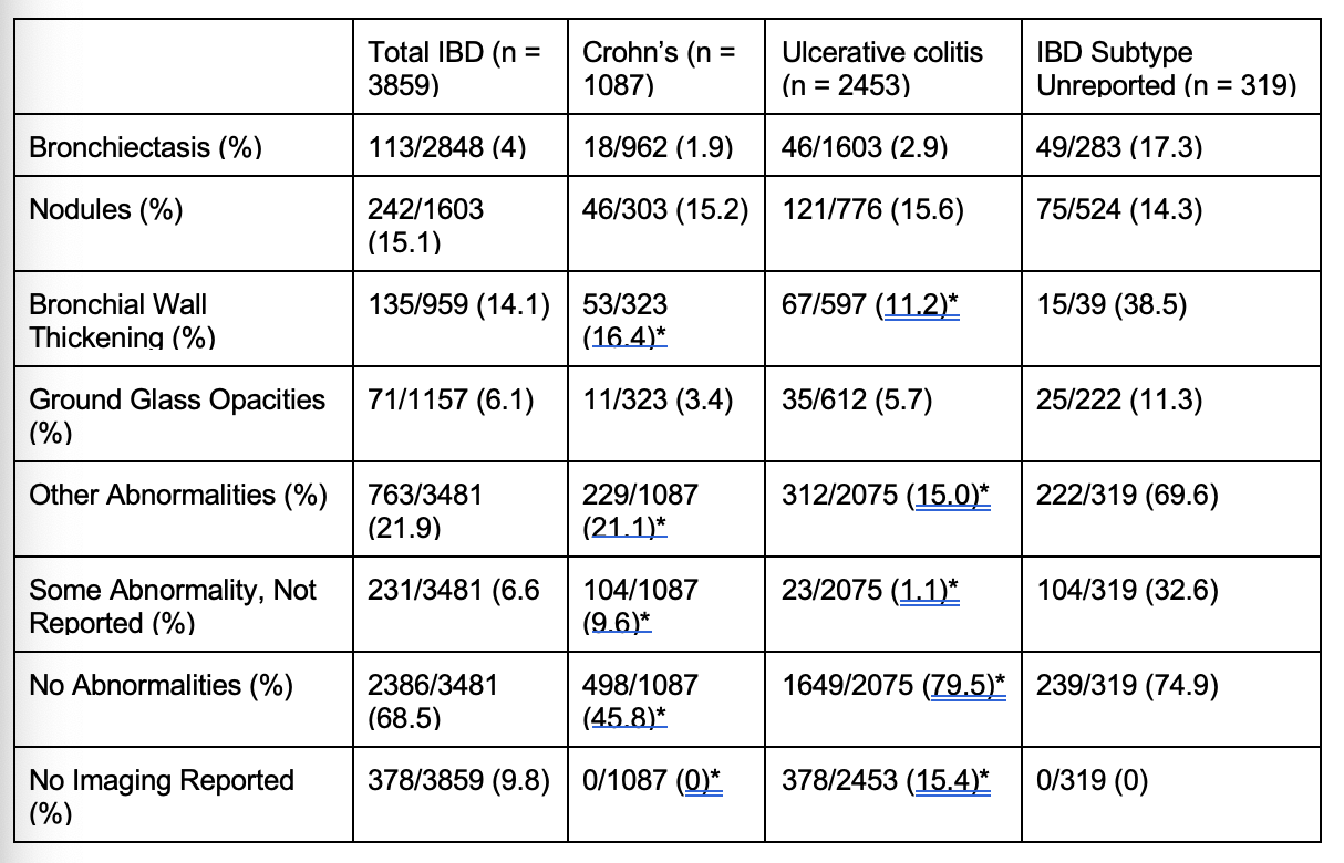
Tatini Mal-Sarkar, MD1, Suruchi Ramanujan, MD2, Varisha Essani, MD3, Joseph Sleiman, MD4, Suha Abushamma, MD4, Taha Qazi, MD4, Florian Rieder, MD4, Katherine Falloon, MD4 1Cleveland Clinic Foundation, Cleveland Heights, OH; 2University of California San Diego, San Diego, CA; 3University of Toledo Medical Center, Toledo, OH; 4Cleveland Clinic Foundation, Cleveland, OH Introduction: Extra-intestinal manifestations (EIMs) affect up to half of all patients with inflammatory bowel disease (IBD), but there is limited literature exploring pulmonary manifestations of IBD. In this systematic review, we evaluated prevalence and pattern of pulmonary abnormalities in patients with IBD who underwent chest imaging. Methods: We conducted a systematic review investigating chest imaging findings in adult IBD patients with and without pulmonary symptoms. Cohort, randomized control trial, case-control, and cross-sectional study designs were eligible for inclusion. We identified 18 studies including 13 cohort and five case-control studies. The primary outcomes were the presence of imaging abnormalities, including bronchiectasis, lung nodules, bronchial wall thickening, and ground-glass opacities. Percentages were taken out of the total number of patients in studies that reported that specific imaging finding. Results: 3859 total IBD patients were included in this study, of whom 1087 (28.2%) were diagnosed with Crohn’s disease (CD) and 2453 (63.6%) were diagnosed with ulcerative colitis (UC). 319 (8.3%) were reported only as IBD (subtype not specified). The most common pulmonary abnormality found on imaging was lung nodules, which affected 242 IBD patients, followed by bronchial wall thickening (n = 135), bronchiectasis (n = 113), then ground glass opacities (n = 71) (Table 1). Bronchial wall thickening was more prevalent among patients with CD than among patients with UC (53 (16.4%) vs 67 (11.2%), p = 0.03). There were no significant differences in prevalence of bronchiectasis, nodules, and ground glass opacities (p = 0.12, p = 0.87, and p = 0.12 respectively). While controls were included in some of the studies examined, only one study reported their chest imaging findings and so comparison of rates of imaging abnormalities between IBD patients and controls was not possible. Discussion: This systematic review found that up to 14.5% of symptomatic and asymptomatic patients with IBD have abnormal chest imaging findings, with the most common findings being nodules, bronchial wall thickening, bronchiectasis, and ground glass opacities. Patients with CD were more likely to have bronchial wall thickening compared to patients with UC. Further research is needed to identify if patients with IBD are at increased risk compared to the general population and which patients with IBD may benefit from additional screening with chest imaging.
Figure: Title: Table 1: Pulmonary Imaging Findings by IBD Diagnosis Caption: IBD = Inflammatory Bowel Disease; CD = Crohn’s Disease; UC = Ulcerative Colitis; NA = Not Applicable; * p < 0.05 compared between IBD diagnoses