Cleveland Clinic Abu Dhabi Abu Dhabi, Abu Dhabi, United Arab Emirates
Doa'a Alkhader, MD, Fatima Alkhyeli, MD, Fatma Mahmoud, MBBCh, Ahmed AlZubaidi, MD, Zaher Koutoubi, MD, Ali Al Jaberi, MD Cleveland Clinic Abu Dhabi, Abu Dhabi, Abu Dhabi, United Arab Emirates Introduction: Gastrointestinal basidiobolomycosis (GIB) is a rare fungal infection in immunocompetent host, characterized by submucosal growth that mimics malignancy and complicates endoscopic diagnosis. Early surgical intervention with surgery and antifungals are critical .
Case Description/
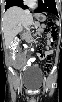
Methods: A 45-year-old man with uncontrolled type 2 diabetes presented with melena, right lower quadrant pain, and weight loss.Eexamination revealed a tender abdominal mass. Labs showed anemia (Hb 67 g/L), leukocytosis (16 x10⁹/L), and eosinophilia. CT revealed a locally invasive 10 cm cecal mass with right sided hydronephrosis [Figure 1]. Colonoscopy demonstrated an ulcerated obstructive, circumferential mass in the ascending colon .Biopsies were inconclusive for malignancy.
After multidisciplinary tumor board review, open right colectomy with ileotransverse anastomosis and diverting loop ileostomy was performed
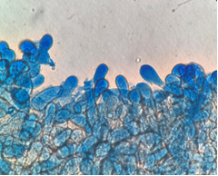
Histology revealed granulomatous inflammation, eosinophilic microabscesses, and pauci-septate hyphae (PAS/GMS+) confirming basidiobolomycosis. [Figure 2]. He was started on amphotericin B followed by oral posaconazole for long-term antifungal therapy. Discussion: GIB caused by Basidiobolus ranarum often mimicsmalignancy or inflammatory conditions,, with hallmark features including abdominal mass, GI bleeding, eosinophilia, and neoplastic-like imaging.Histologic clues (granulomatous inflammation,Splendore-Hoeppli phenomenon). with culture stains confirm diagnosis. This case underscores the diagnostic challenge of GIB despite classic presentation. Multidisciplinary collaboration and histopathologic analysis prevented delayed treatment. Combined surgical resection and prolonged antifungals remain essential for favorable outcomes..
Figure: Figure 1. Coronal section of Computed tomography (CT) of the abdomen and pelvis demonstrating a bulky cecal mass measuring 10 x 7 x 10 cm, with lymphovascular infiltration, lateral invasion into the transverse fascia and muscle, and medial infiltration to the right ureter, causing mild hydronephrosis
Figure: Figure 2. Microorganisms with fungal-like features including pauci-septate hyphae with surrounding eosinophilic cuffing by periodic acid-schiff (PAS) and Gomori methenamine silver (GMS) staining.
Disclosures: Doa'a Alkhader indicated no relevant financial relationships. Fatima Alkhyeli indicated no relevant financial relationships. Fatma Mahmoud indicated no relevant financial relationships. Ahmed AlZubaidi indicated no relevant financial relationships. Zaher Koutoubi indicated no relevant financial relationships. Ali Al Jaberi indicated no relevant financial relationships.
Doa'a Alkhader, MD, Fatima Alkhyeli, MD, Fatma Mahmoud, MBBCh, Ahmed AlZubaidi, MD, Zaher Koutoubi, MD, Ali Al Jaberi, MD. P1321 - Fungal Impostor: Gastrointestinal Basidiobolomycosis Mimicking an Obstructive Colon Mass, ACG 2025 Annual Scientific Meeting Abstracts. Phoenix, AZ: American College of Gastroenterology.


 photo")