University of Arkansas for Medical Sciences Little Rock, AR
Gomathy Nageswaran, MBBS1, Rupinder Mann, MD2, Sumant Inamdar, MD1 1University of Arkansas for Medical Sciences, Little Rock, AR; 2University of Arkansas, Little Rock, AR Introduction: Fistulas between the extrahepatic biliary system and intestinal organs are rare, mainly secondary to choledocholithiasis, and rarely arise from the stomach (1). Here we present what we believe to be the first reported case of endoscopic stenting to treat a gastro-biliary fistula secondary to choledocholithiasis.
Case Description/
Methods: A 35-year-old man with a history of hypertension, anxiety disorder, psoriasis, and alcoholic liver disease presented to the hospital for abdominal pain and nausea. Physical exam at presentation was normal except for diffuse abdominal tenderness. Laboratory workup showed elevated white cell count to 12.6 K/µL, a total bilirubin of 13.5 mg/dL, direct bilirubin of 8.7 mg/dL, GGT of 337 IU/L, SGOT/SGPT of 126/133 U/L, ALP of 101 U/L. On endoscopic ultrasound, the common bile duct (CBD) was dilated to 15.5 mm with stones. An endoscopic retrograde cholangiopancreaticography (ERCP) was performed. The cholangiogram showed a biliary stone and a communication between the CBD and the stomach, a gastro-biliary fistula. Spyglass cholangioscopy was performed with the removal of the CBD stone and visualization of the fistula. A plastic biliary stent was placed into the CBD. On repeat ERCP after three months, the stent was removed, and the occlusion cholangiogram was normal. Discussion: Common causes of spontaneous fistula include longstanding, untreated choledocholithiasis, recurrent cholangitis, peptic ulcer disease, and rarely neoplasms(2). Endoscopic options for an extra-biliary fistula include either sphincterotomy or stenting. Both techniques increase the transpapillary biliary flow, thereby reducing the leak; however, stenting is usually preferred in younger patients to preserve the sphincter (2,3). Stents can be of two types – plastic or metal, and they are generally left in place for about 2-3 months for leak occlusion. Surgical repair is usually reserved for the drainage of large collections or recurrent leaks (3).
References:
1. Crespi M, Montecamozzo G, Foschi D. Diagnosis and Treatment of Biliary Fistulas in the Laparoscopic Era. Gastroenterol Res Pract. 2016;2016:1–6.
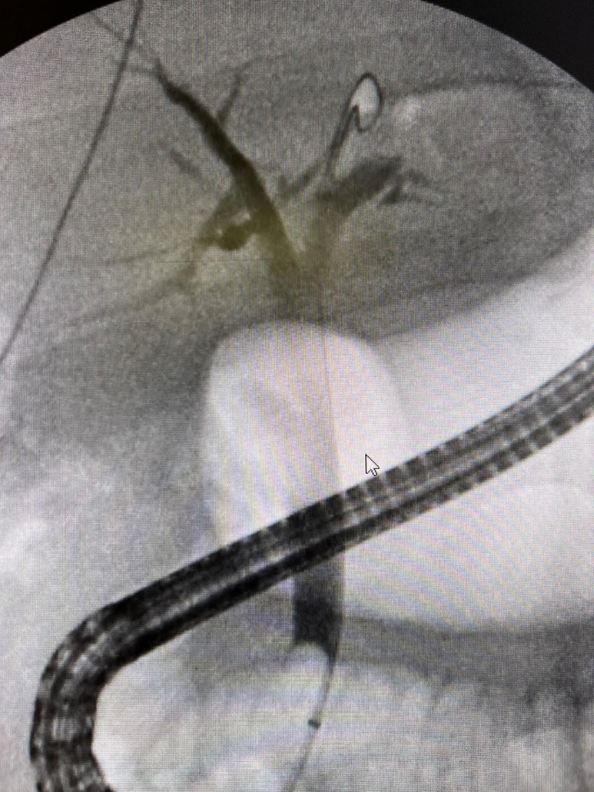
Figure: Figure 1: Fluoroscopic image obtained during endoscopic retrograde cholangiopancreatography (ERCP) showing successful positioning of a plastic stent as a means for occlusion of a gastro-biliary fistula
Disclosures: Gomathy Nageswaran indicated no relevant financial relationships. Rupinder Mann indicated no relevant financial relationships. Sumant Inamdar indicated no relevant financial relationships.
Gomathy Nageswaran, MBBS1, Rupinder Mann, MD2, Sumant Inamdar, MD1. P1468 - Looking Through the Spyglass: Stenting of a Gastro-biliary Fistula Secondary to Choledocholithiasis, ACG 2025 Annual Scientific Meeting Abstracts. Phoenix, AZ: American College of Gastroenterology.