Maxwell Charlat, MD, Makeda Dawkins, MD, Shireen Pais, MD Westchester Medical Center, Valhalla, NY Introduction: Lumen-apposing metal stents (LAMS) have emerged as a minimally invasive option for creating drainage tracts between gastrointestinal structures. They are increasingly used for the treatment of large, well-encapsulated pancreatic pseudocysts and walled-off necroses. We present a case of a large peripancreatic fluid collection initially considered for LAMS, highlighting the need for thorough diagnostic evaluation before proceeding with endoscopic drainage.
Case Description/
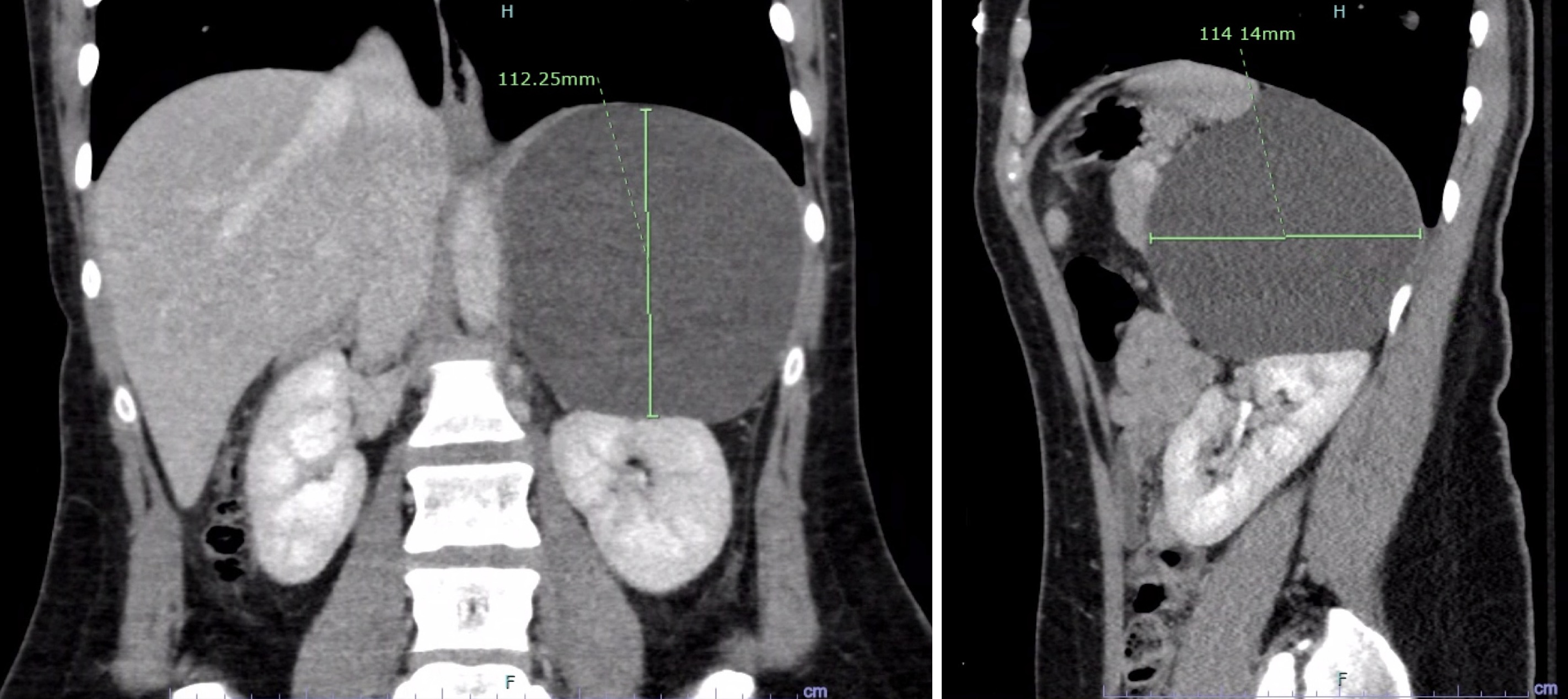
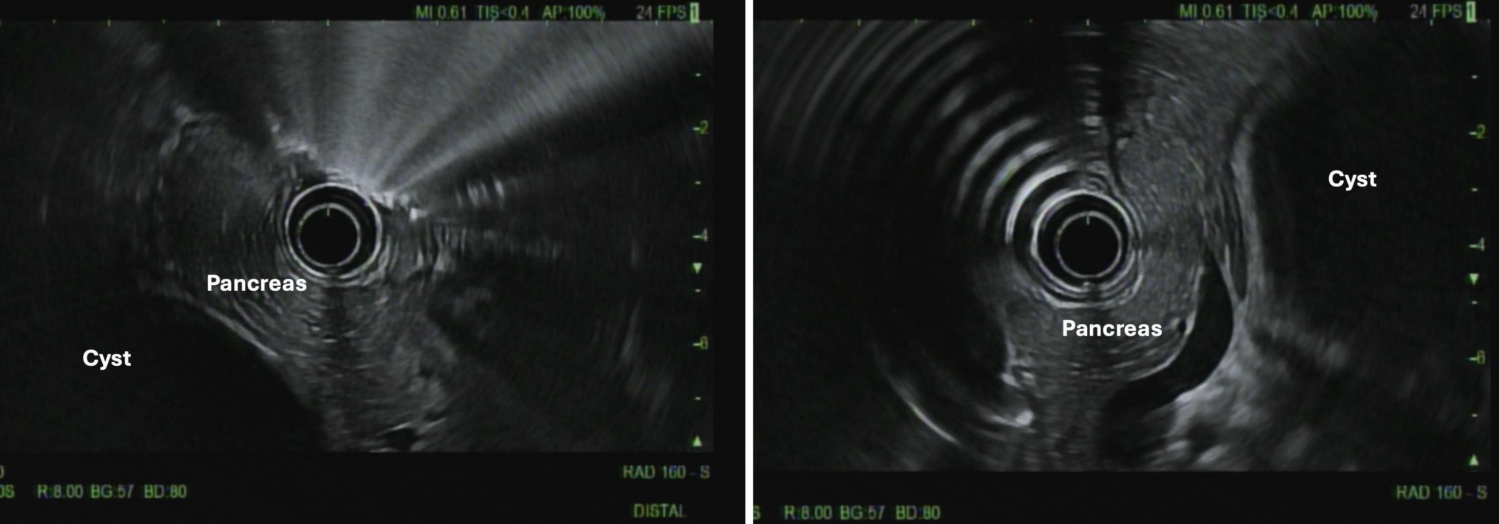
Methods: A 24-year-old woman with no significant medical history presented to the emergency department after a motor vehicle collision. Imaging revealed no traumatic injury but showed an 11.1 × 11.2 × 11.4 cm unilocular cyst adjacent to the pancreatic tail, spleen, and upper left renal pole (Figure 1). On further history, she reported three years of intermittent left-sided abdominal pain with emesis and denied any prior gastrointestinal disease. Endoscopic ultrasound demonstrated a normal pancreas and a 14 × 9 cm extraluminal cyst in the pancreatic tail (Figure 2). Fine needle aspiration was performed. Post-procedurally, the patient experienced worsening abdominal pain, and LAMS placement was considered. However, fluid analysis revealed amylase 44 U/L, carcinoembryonic antigen 0.4 ng/mL, glucose 94 mg/dL, and negative cytology—findings inconsistent with a pancreatic pseudocyst or mucinous lesion. A mesenchymal cyst was suspected. Upon multidisciplinary review with oncologic surgery, operative management was recommended. Intraoperatively, the cyst was found in the retroperitoneum, posterior to the spleen, consistent with a mesenteric cyst. Laparoscopic excision was completed, and the patient’s symptoms resolved. Discussion: Mesenteric cysts are rare benign intra-abdominal lesions, thought to arise from ectopic lymphatic tissue, with an incidence of 4–10 per million hospital admissions. Though LAMS was technically feasible, drainage alone carries a risk of mesenteric cyst recurrence and complications such as rupture, volvulus, and obstruction. This case highlights the critical importance of balancing enthusiasm for LAMS and other minimally invasive endoscopic procedures with the imperative to minimize patient harm and avoid repeat interventions.
Figure: Figure 1. CT images of the abdomen showed a large cyst in the coronal (left) and sagittal (right) planes.
Figure: Figure 2. Endoscopic ultrasound (EUS) showed an extraluminal cyst in the pancreatic tail.
Disclosures: Maxwell Charlat indicated no relevant financial relationships. Makeda Dawkins indicated no relevant financial relationships. Shireen Pais indicated no relevant financial relationships.
Maxwell Charlat, MD, Makeda Dawkins, MD, Shireen Pais, MD. P1465 - Not Every Cyst Needs a Lumen-Apposing Metal Stent: Mesenteric Cyst Mimicking Peripancreatic Fluid Collection, ACG 2025 Annual Scientific Meeting Abstracts. Phoenix, AZ: American College of Gastroenterology.

.jpg "Maxwell Charlat, MD photo")