Ariba Khan, MD1, Bakr Alhayek, MD1, Michael Dennis. Harris, MD2 1AdventHealth, Tampa, FL; 2ADVENTHEALTH TAMPA, Tampa, FL Introduction: Mucinous cystic neoplasms of the liver (MCN-L) are a rare subset of cystic hepatic tumors representing less than 5% of hepatic cysts. They occur exclusively in middle-aged females and carry a malignant potential, having been historically termed biliary cystadenoma/cystadenocarcinoma. MCN-L usually arises within the liver parenchyma and, less frequently, involves the extrahepatic bile ducts. Here we discuss a rare presentation of MCN-L manifesting as obstructive jaundice.
Case Description/
Methods: A 22-year-old previously healthy female with a 5-year history of intermittent abdominal pain, nausea, and vomiting was transferred to our tertiary-care hospital for acute worsening of symptoms along with clinical signs of obstructive jaundice. A cholestatic pattern of liver enzyme elevation was noted (T. Bilirubin - 2.9 mg/dL, ALP - 293 U/L). Initial abdominal CT revealed a saccular cystic lesion in the caudate lobe of the liver, along with mild intrahepatic ductal dilation, secondary to a focal obstruction/stricture of the common hepatic duct as seen on MRCP. A presumptive diagnosis of a possible congenital anomaly was being considered. EUS/ERCP with cholangioscopy was performed, demonstrating a soft tissue mass in the common hepatic duct and prominent blood vessels, worrisome for a neoplastic process. Pathology taken from cholangioscopy confirmed a low-grade mucinous cystic neoplasm. Ultimately, the patient underwent a robotic extrahepatic biliary resection, liver resection, and a Roux-en-Y hepaticojejunostomy. Histopathology revealed a mucinous cyst lined by columnar epithelium overlying estrogen receptor-positive ovarian-type stroma, consistent with a low-grade MCN; no invasive carcinoma was identified. Discussion: Due to insufficient recognition and the absence of distinct clinical and radiological features, MCN-Ls are often misdiagnosed, resulting in a delay in treatment. Evaluation of a multi-loculated hepatic cyst in a female patient should begin with high-resolution imaging, supplemented with tumor markers, cholangioscopy for tissue acquisition, and definitive intraductal characterization. A review of 22 adult cases (2015–2025) found that all recurrences followed non-curative procedures, while patients undergoing primary margin-negative resection remained disease-free. These findings underscore that definitive treatment requires formal anatomic hepatectomy with concomitant bile duct resection when indicated, with long-term outcomes determined by surgical adequacy rather than tumor biology.
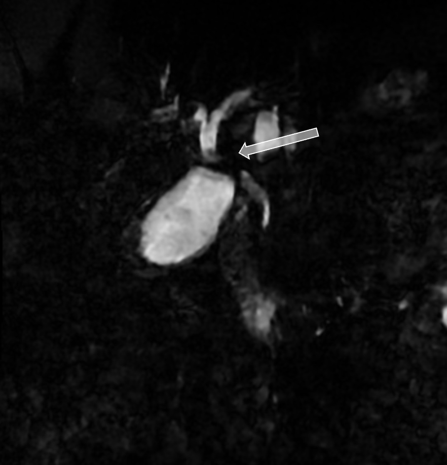
Figure: MRCP showing focal hilar stricture of the common hepatic duct with mild upstream biliary duct dilation.
Figure: Left image: Cholangioscopy showing a well-circumscribed common bile duct (CBD) mass. Right image: Estrogen receptor (ER) staining showing nuclear positivity in spindle cells, suggesting ovarian-type stroma, with negative staining in the single-layer surface epithelial lining on intra-operative pathology.
Disclosures: Ariba Khan indicated no relevant financial relationships. Bakr Alhayek indicated no relevant financial relationships. Michael Harris indicated no relevant financial relationships.
Ariba Khan, MD1, Bakr Alhayek, MD1, Michael Dennis. Harris, MD2. P1711 - An Unusual Tumor in an Uncommon Patient: Hepatic Mucinous Cystic Neoplasm Causing Biliary Obstruction, ACG 2025 Annual Scientific Meeting Abstracts. Phoenix, AZ: American College of Gastroenterology.


.jpg "Ariba Khan, MD (she/her/hers) photo")