Samra Iqbal, MD1, Sushil Sharma, MD2, Aram Aslanian, MD2, Jairah Shaikh, MD2, Adel Iqbal, BA3 1Texas Tech University Health Sciences Center, Helotes, TX; 2Texas Tech University Health Sciences Center, Amarillo, TX; 3Rice University, San Antonio, TX Introduction: Acute appendicitis is among the most common causes of emergency abdominal surgery, affecting up to 7–8% of the population. While typically caused by luminal obstruction and inflammation, a small percentage of cases may be due to underlying neoplasms. Mucinous adenocarcinoma of the appendix is a rare malignancy, accounting for less than 0.5% of gastrointestinal cancers. It often presents with symptoms indistinguishable from uncomplicated appendicitis, posing a diagnostic challenge. Preoperative imaging may not reveal the tumor, and diagnosis is often made postoperatively via histopathological evaluation. Early detection is crucial, as it influences staging, prognosis, and management. We present a rare case of invasive mucinous adenocarcinoma presenting as routine appendicitis.
Case Description/
Methods: A 58-year-old male with no significant medical history presented with 48 hours of progressive right lower quadrant pain, nausea, and vomiting. The pain initially began periumbilically before localizing to the right lower quadrant. He denied fever or changes in bowel habits. Physical exam revealed tenderness, guarding, and rebound in the right lower quadrant. Labs showed mild leukocytosis (12,500/μL). CT abdomen/pelvis with contrast showed a dilated appendix with periappendiceal fat stranding, consistent with acute appendicitis. No mass or lymphadenopathy was seen. The patient underwent laparoscopic appendectomy. Intraoperatively, the appendix appeared enlarged but without visible mass or perforation. Postoperative recovery was uneventful. Histopathology revealed invasive mucinous adenocarcinoma with mucin pools and glandular structures invading through the appendiceal wall. Immunohistochemistry confirmed the diagnosis. The patient was referred to oncology for further staging and management. Discussion: This case highlights a rare but important diagnostic consideration in patients presenting with appendicitis, particularly those over 50. Appendiceal mucinous adenocarcinoma can mimic typical appendicitis and evade preoperative detection. Routine histopathologic analysis of appendectomy specimens is critical for timely diagnosis. Early identification allows for appropriate oncologic workup and may necessitate right hemicolectomy or surveillance. Clinicians should consider malignancy in atypical or persistent appendicitis presentations, even when imaging suggests a benign process.
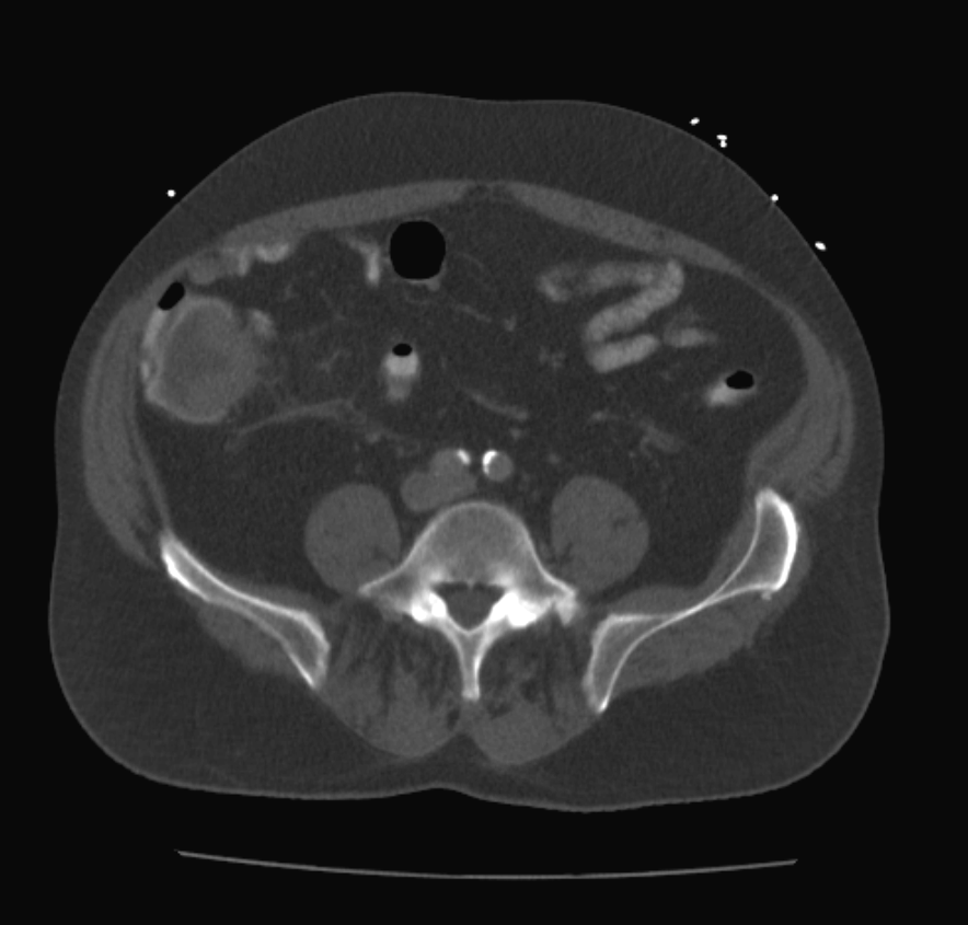
Figure: Figure 1. Axial contrast-enhanced CT scan of the abdomen showing an enlarged, fluid-filled appendix with surrounding fat stranding, findings consistent with acute appendicitis.
Figure: Figure 2. Histopathologic section of the appendix showing invasive mucinous adenocarcinoma.
Disclosures: Samra Iqbal indicated no relevant financial relationships. Sushil Sharma indicated no relevant financial relationships. Aram Aslanian indicated no relevant financial relationships. Jairah Shaikh indicated no relevant financial relationships. Adel Iqbal indicated no relevant financial relationships.
Samra Iqbal, MD1, Sushil Sharma, MD2, Aram Aslanian, MD2, Jairah Shaikh, MD2, Adel Iqbal, BA3. P1940 - Invasive Mucinous Adenocarcinoma of the Appendix Masquerading as Acute Appendicitis: A Rare Diagnostic Challenge, ACG 2025 Annual Scientific Meeting Abstracts. Phoenix, AZ: American College of Gastroenterology.