Cook County Health and Hospital Systems Chicago, IL
Patricia Zarza, MD1, Robert Kwei Nsoro, MD1, Daniel Guifarro Rivera, MD1, Alejandro Nieto Dominguez, MD1, Ahad Samad, MD2 1Cook County Health and Hospital Systems, Chicago, IL; 2Insight Hospital & Medical Center, Chicago, IL Introduction: We present a case of epidermoid metaplasia, a rare condition documented only in case reports and case series. It was first described in 1997 by Nakanishi et al. in a 58-year-old male with esophageal cancer. Histological findings were described as unusual epithelial changes and esophageal mucosal epidermization, consistent with what is currently described as epidermoid metaplasia.
Case Description/
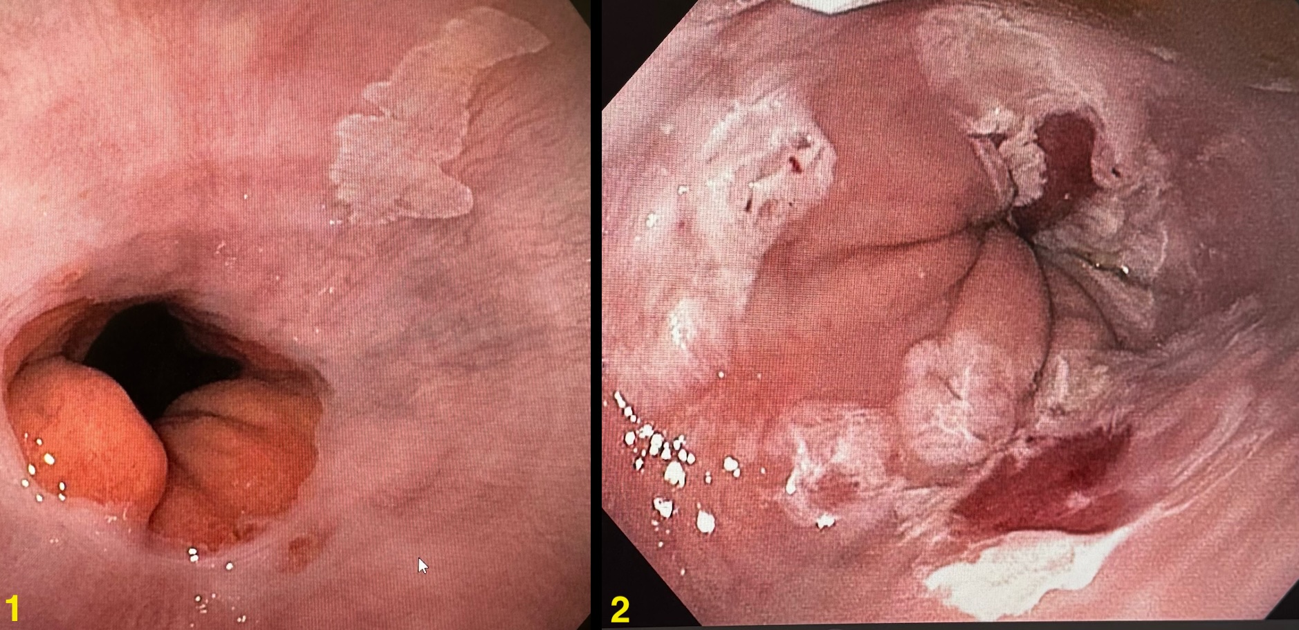
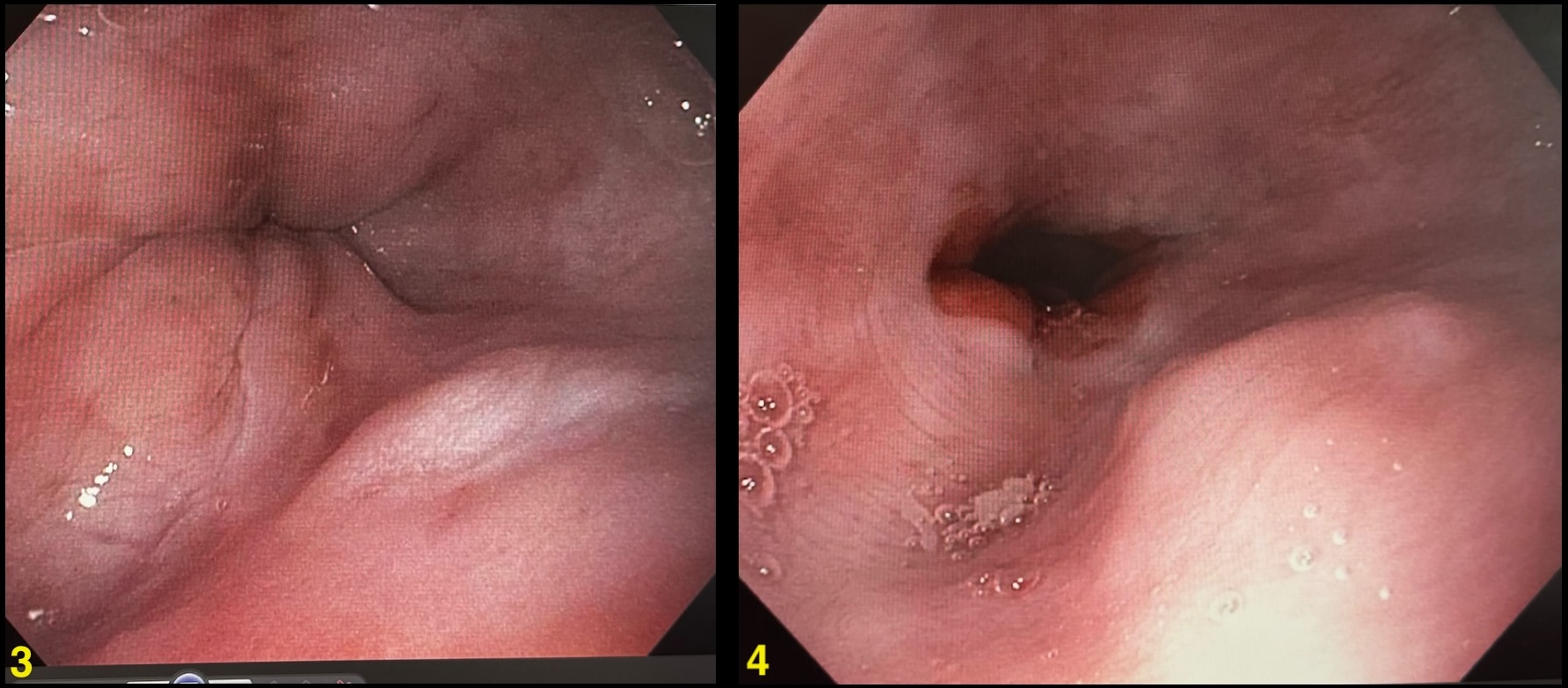
Methods: 50-year-old male with history of hypertension, prediabetes, obesity, alcohol and tobacco use disorder who presented with a one-year history of solid dysphagia and globus sensation. He denied reflux-type symptoms, weight loss, or hematemesis. Physical exam was unremarkable and laboratory did not show anemia. Endoscopic evaluation revealed sharply demarcated white-to-tan mucosal plaques in the mid-to-distal esophagus. Subsequent biopsy confirmed epidermoid metaplasia. The patient underwent radiofrequency ablation and was scheduled for endoscopic surveillance at six-month intervals. Discussion: Epidermoid metaplasia has been described mainly in the middle-aged and elderly population. In a case series of 18 patients by Singh et al., the age range at diagnosis was 47 to 87 years, with a mean age of 68.2 and a slight male predominance (56%). In a larger case series in a tertiary center conducted by Cotteau et al., among 1048 esophageal samples, epidermoid metaplasia was identified in only two cases. The etiology remains uncertain, though chronic exposure to alcohol and tobacco appears to play a significant role. Targeted next-generation sequencing has demonstrated TP53 mutations in 56% of cases, alongside alterations in EGFR, HRAS, and PIK3CA, suggesting a potential predisposition to neoplastic transformation. Epidermoid metaplasia is often identified in patients undergoing endoscopy for dysphagia, GERD, or surveillance for a history of esophageal cancer or Barrett's esophagus. Endoscopically, it is often described as a sharply demarcated patch or plaque consisting of white-to-tan mucosa. The gold standard for diagnosis is histopathological evaluation revealing esophageal squamous mucosa with hyperorthokeratosis. Currently, there are no standardized treatment guidelines for this condition. Management options include endoscopic surveillance, radiofrequency ablation, and endoscopic mucosal resection. While the pathogenesis remains incompletely understood, early identification and appropriate surveillance strategies are essential for mitigating the potential progression to squamous neoplasia.
Figure: 1. December 2023 EGD 2. September 2024 RFA
Figure: 3. December 2024 EGD 4. February 2025 EGD
Disclosures: Patricia Zarza indicated no relevant financial relationships. Robert Kwei Nsoro indicated no relevant financial relationships. Daniel Guifarro Rivera indicated no relevant financial relationships. Alejandro Nieto Dominguez indicated no relevant financial relationships. Ahad Samad indicated no relevant financial relationships.
Patricia Zarza, MD1, Robert Kwei Nsoro, MD1, Daniel Guifarro Rivera, MD1, Alejandro Nieto Dominguez, MD1, Ahad Samad, MD2. P2826 - A Rare Case of Epidermoid Metaplasia in the Esophagus, ACG 2025 Annual Scientific Meeting Abstracts. Phoenix, AZ: American College of Gastroenterology.