Universidad Central del Caribe School of Medicine Tampa, FL
Jerry Cruz Rodriguez, BS1, Karolane A. González González, BS2, Camila I. Delgado Gonzalez, 3, Diego J. Diaz Mayor, MD4, Aura F. Delgado Cifuentes, MD5, Jorge Billoch, MD6, Francis Bauldrick, MD7 1Universidad Central del Caribe School of Medicine, Bayamon, Puerto Rico; 2Universidad Central del Caribe School of Medicine, Guaynabo, Puerto Rico; 3Universidad de Puerto Rico, Bayamon, Puerto Rico; 4VA Caribbean Healthcare System, San Juan, Puerto Rico; 5Hospital Metropolitano, San Juan, Puerto Rico; 6HRP Labs, San Juan, Puerto Rico; 7Auxilio Mutuo Hospital, San Juan, Puerto Rico Introduction: Mesenteric fibromatosis (MF) is a benign form of desmoid tumors that originates in the abdominal mesentery. Although benign, MF often demonstrates locally aggressive progression. It is characterized by the proliferation of well-differentiated fibroblasts and myofibroblasts that, as they grow, may compress surrounding organs, leading to intestinal obstruction, ischemia, and perforation. Accounting for two to four new cases per million people per year and with an incidence of only 0.03%, MF is considered a rare tumor. In the abdomen, apart from developing within the mesentery, it may also be seen in the mesocolon and the retroperitoneum. We report a case with mild, nonspecific symptoms of MF that required a high index of suspicion to perform the appropriate workup and extraction.
Case Description/
Methods: A 24-year-old woman with no significant medical history arrived at the GI clinic after experiencing nausea, constipation, and abdominal pain for six months. MRI and ultrasound of the abdomen revealed a large 3.3 × 3.7 cm mass near the splenic hilum with evidence of colonic and pancreatic tail compression and no invasion. Subsequent colonoscopy revealed an extracolonic mass with significant compression of the colon. EUS with biopsy was remarkable for fibroblastic/myofibroblastic cell proliferation of unknown significance, characteristic of primary MF. Exploratory laparotomy revealed a densely fibrous mass adhered to the splenic hilum, splenic flexure of the colon, and pancreatic tail, which required splenectomy, distal pancreatectomy, and partial colectomy. She tolerated the procedure without complications, and no recurrence occurred during screening. Discussion: MF commonly occurs in women of reproductive age, either associated with another condition, such as Gardner’s syndrome or as a spontaneous isolated mesenteric fibromatosis (SIMF). It has been previously associated with past abdominal surgeries or estrogen exposure. Despite its benign nature, it can grow aggressively and cause mass effect complications on local organs. Abdominal imaging with the US, CT scan, or MRI can detect extraintestinal lesions, but a colonoscopy must also be performed due to the possible presence of intracolonic tumors and its feasibility of obtaining a biopsy for diagnosis. Surgical resection is the first choice for management. Yearly follow-up is recommended in all patients due to its recurrence of 23 to 50%.
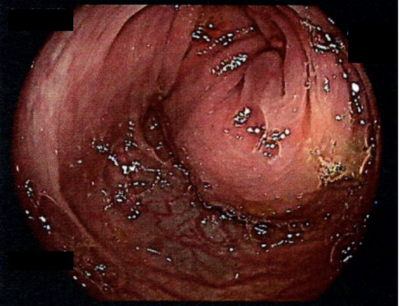
Figure: Colonoscopic view of an extracolonic mesenteric fibromatosis mass causing significant compression of the colonic lumen.
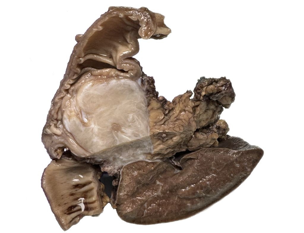
Figure: Postoperative view of a mesenteric fibromatosis mass attached to the pancreatic tail, colon, and splenic hilum.
Disclosures: Jerry Cruz Rodriguez indicated no relevant financial relationships. Karolane González González indicated no relevant financial relationships. Camila Delgado Gonzalez indicated no relevant financial relationships. Diego Diaz Mayor indicated no relevant financial relationships. Aura Delgado Cifuentes indicated no relevant financial relationships. Jorge Billoch indicated no relevant financial relationships. Francis Bauldrick indicated no relevant financial relationships.
Jerry Cruz Rodriguez, BS1, Karolane A. González González, BS2, Camila I. Delgado Gonzalez, 3, Diego J. Diaz Mayor, MD4, Aura F. Delgado Cifuentes, MD5, Jorge Billoch, MD6, Francis Bauldrick, MD7. P2582 - Primary Mesenteric Fibromatosis With Multi-Organ Compression: A Case of Surgical Complexity, ACG 2025 Annual Scientific Meeting Abstracts. Phoenix, AZ: American College of Gastroenterology.

 photo")