Wellstar Health System - Cobb Medical Center Austell, GA
Jill Bhavsar, MD, Manjusha Das, MD Wellstar Health System - Cobb Medical Center, Austell, GA Introduction: Solid pseudopapillary neoplasm is a rare pancreatic tumor characterized by low malignant potential and unique pathological features. Despite being classified as malignant, this tumor's growth is indolent and complete surgical resection yields excellent prognosis. Most commonly found in females in their 2nd or 3rd decade of their life; the presentation of this tumor is insidious and often discovered incidentally.
Case Description/
Methods: A 40 year old female was admitted after an episode of large volume hematemesis and melena. She reportedly had no family history of malignancies, no recent use of ibuprofen, no prior endoscopies. CT imaging showed no active bleeding but a possible splenic vein thrombosis with prominent collateral vessels concerning possible variceal bleeding. She had recurrence of projectile large volume hematemesis and became hemodynamically unstable. In the emergent esophagogastroduodenoscopy (EGD), she was noted to have type 1 isolated gastric varices in gastric fundus with high-risk stigmata of recurrent bleeding. Coil embolization of left gastric and accessory left gastric artery was performed by Interventional Radiology (IR). As definitive treatment, she underwent laparoscopic distal pancreatectomy with splenectomy. Its pathology report identified solid pseudopapillary neoplasm confined to the pancreatic tail with negative surgical margins. She did well on follow up visits and was recommended repeat imaging in 12 months with no other adjuvant treatment required. Discussion: Pancreatic solid pseudopapillary neoplasm accounts for 2 to 3% of all pancreatic neoplasms and 0.9% to 2.7% of exocrine pancreatic neoplasms. In 85% of cases, the mass is limited to pancreas while in 10-15% of cases present with metastasis. Most of the patients are asymptomatic, but some have non specific and vague symptoms like abdominal pain, discomfort, nausea, vomiting and early satiety. Very rarely some patients present with jaundice or acute hemoperitoneum due to tumor rupture. It presents as well circumscribed, encapsulated and heterogeneous with hemorrhagic and cystic degeneration on imaging but endoscopic ultrasound guided fine needle aspiration may help establish the diagnosis. The treatment of choice is complete resection if possible, unresectable cases have shown promising responses with radiotherapy, chemotherapy and hormonal therapy and patients have survival rates above 90% after treatment.
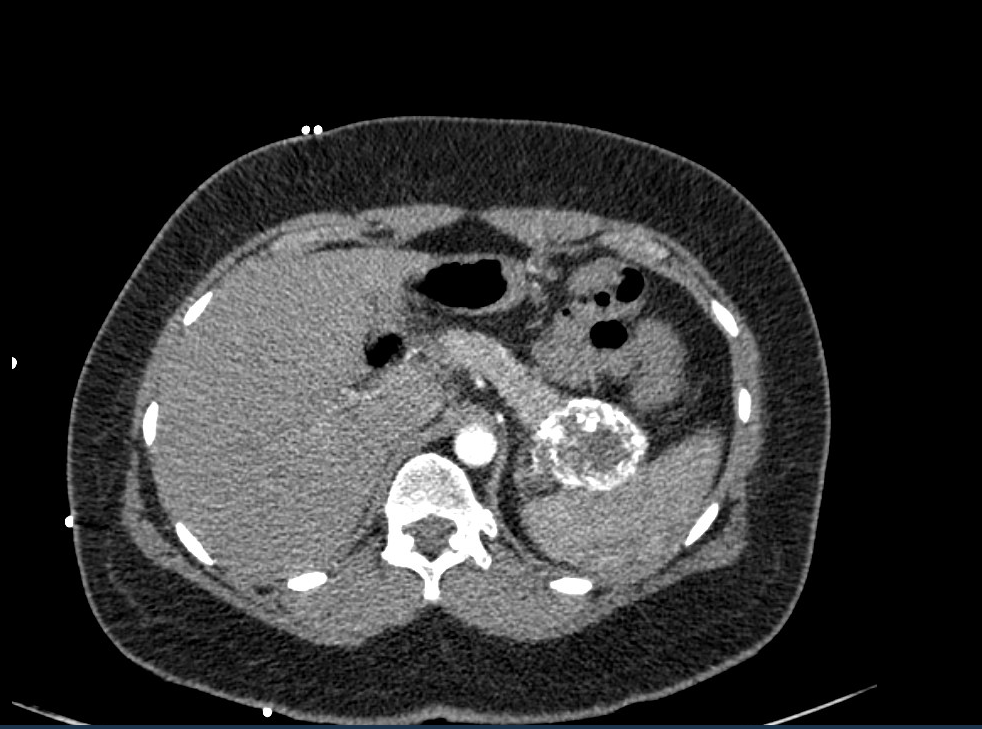
Figure: Large peripherally calcified hypoattenuating lesion within or abutting the pancreatic tail.
Disclosures: Jill Bhavsar indicated no relevant financial relationships. Manjusha Das indicated no relevant financial relationships.
Jill Bhavsar, MD, Manjusha Das, MD. P2272 - Acute Gastrointestinal Bleeding: A Rare Presentation of Solid Pseudopapillary Neoplasm of Pancreas - A Case Report, ACG 2025 Annual Scientific Meeting Abstracts. Phoenix, AZ: American College of Gastroenterology.

 photo")