University of Texas Health San Antonio San Antonio, TX
Award: ACG Presidential Poster Award
Parth Patel, MD, MBA1, Adam Vinall, MD1, Umesh Boregowda, MD1, Shuhaib Ali, MD1, Apeksha Agarwal, MD1, Courtney Thomas, DO1, Patrick Snyder, MD2, Alexis Pavle, MD3 1University of Texas Health San Antonio, San Antonio, TX; 2San Antonio Military Medical Center, San Antonio, TX; 3University of Texas Health Science Center at San Antonio, San Antonio, TX Introduction: Li-Fraumeni syndrome (LFS) is a rare autosomal dominant disorder that greatly increases the risk of adrenocortical carcinoma, breast cancer, central nervous system tumors, osteosarcoma, and soft tissue sarcomas. Leiomyosarcoma (LMS) is an aggressive soft tissue sarcoma arising from smooth muscle cells, most often in the uterus, gastrointestinal tract, and blood vessels. Gastric adenocarcinoma is also linked to LFS, with expert guidelines recommending screening endoscopy every 2–3 years starting at age 25. Here, we present the first documented case of gastric leiomyosarcoma in an individual with Li-Fraumeni syndrome.
Case Description/
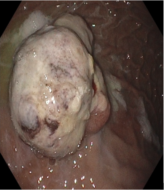
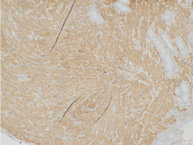
Methods: A 27-year-old male with a past medical history of Li-Fraumeni Syndrome, complicated by acute lymphoblastic leukemia, status post chemotherapy, radiotherapy, and bone marrow transplant at age 14, presented with shortness of breath and lightheadedness. He endorsed episodes of intermittent dark stool and epigastric tenderness which improved with food. He has an extensive family history of cancer in his immediate family. Initial hemoglobin was 7.3 g/dL, significantly lower than his baseline of 14.0 g/dL six months prior. A 3 phase CT abdomen and pelvis showed no acute intra-abdominal abnormality. Upper endoscopy revealed a large, exophytic mass measuring approximately 5 to 6 cm, with oozing bleeding, in the gastric body along the greater curvature (Image 1). Biopsy of the mass revealed a high-grade leiomyosarcoma (Image 2). Immuno-histological staining showed neoplastic cells expressing smooth muscle actin (SMA) and desmin. The patient underwent a partial gastrectomy, and pathology revealed a 10 x 7 x 2 cm leiomyosarcoma with negative margins. However, five months later, follow-up scans showed tumor recurrence, now measuring 18 x 18 x 14 cm. He began chemotherapy with ifosfamide and doxorubicin, along with radiotherapy, but his condition continued to decline, and he was transitioned to hospice care. Discussion: LFS is a highly penetrant hereditary cancer predisposition syndrome primarily driven by germline TP53 mutations with rare gastric involvement. To our knowledge, this is the first documented case of gastric leiomyosarcoma in a LFS patient. This case expands the spectrum of malignancies associated with LFS and underscores the importance of considering rare tumor types in patients with hereditary cancer syndromes. Further research is needed to evaluate screening and surveillance strategies for early detection and treatment of these rare malignancies.
Figure: Large, exophytic mass measuring approximately 5 to 6 cm, with oozing and bleeding in the gastric body
Disclosures: Parth Patel indicated no relevant financial relationships. Adam Vinall indicated no relevant financial relationships. Umesh Boregowda indicated no relevant financial relationships. Shuhaib Ali indicated no relevant financial relationships. Apeksha Agarwal indicated no relevant financial relationships. Courtney Thomas indicated no relevant financial relationships. Patrick Snyder indicated no relevant financial relationships. Alexis Pavle indicated no relevant financial relationships.
Parth Patel, MD, MBA1, Adam Vinall, MD1, Umesh Boregowda, MD1, Shuhaib Ali, MD1, Apeksha Agarwal, MD1, Courtney Thomas, DO1, Patrick Snyder, MD2, Alexis Pavle, MD3. P2140 - Gastric Leiomyosarcoma in a Patient With Li-Fraumeni Syndrome, ACG 2025 Annual Scientific Meeting Abstracts. Phoenix, AZ: American College of Gastroenterology.