Monmouth Medical Center, Robert Wood Johnson Medical School of Rutgers University Long Branch, NJ
Karan J.. Yagnik, MD, Suryansh Atreya, MD, Pranay Joshi, MD, Sankalp Acharya, MD, Dhramesh Kaswala, MD, Wael Ghali, MD Monmouth Medical Center, Robert Wood Johnson Medical School of Rutgers University, Long Branch, NJ Introduction: We describe a rare case of acute abdominal pain initially misdiagnosed as acute cholecystitis, which was ultimately found to be gastric band erosion occurring 20 years after LAGB placement, complicated by abscess formation along the epithelialized tract.
Case Description/
Methods: A female in her 60s came to the emergency department (ED) complaining of severe abdominal pain, fever and chills. Patient has a past history of LAGB placement 20 years ago. Upon arrival, she was vitally stable. Physical examination showed the epigastric and RUQ tenderness. Labs showed WBC 22.6 x 10*3 u/L with 90% neutrophilia. CT abdomen/pelvis showed unremarkable gastric band. The US abdomen showed nonspecific gallbladder wall thickening. Patient was managed for acute cholecystitis.
Next day, her labs showed CRP 308 mg/L, ESR >130 mm/hr, WBCs 20.8 x 10*3 u/L. HIDA scan was normal. Due to worsening abdominal pain EGD was performed showing band erosion into the stomach. Laparoscopy revealed the band erosion of 50% into the stomach, intra-abdominal abscess throughout the epithelized tubing track. The band was removed, abscesses were drained, and a partial gastrectomy was performed.
Next day, she had worsening RUQ pain. Upper GI series showed leak at lesser curvature, managed conservatively. Following day, she spiked a fever with WBC increased to 25 x 10*3 u/L. CT abdomen/pelvis with IV contrast showed no contrast leak, no evidence of drainable collection, but some fluid in the right upper abdominal wall. Bedside I&D yeilded purulent drainage. Patient improved thereafter. Discussion: Highlight this case is gastric band erosion after 20 years of LAGB placement. It is frequently associated with the cessation of weight loss, and progressive weight regain. However, this symptom is often attributed to late procedural failure—an interpretation that may lead clinicians to under-look erosion as a possible cause due to it occurring relatively earlier in the course.
Another takeaway point in our study is that CT scan failed to show band erosion and diagnosis of band erosion is made through direct exploration by endoscopy. CT scan is sensitive (88%) to diagnose gastric band erosion, endoscopy is gold standard (100%).
We emphasize here that band erosion and other complications of LAGB must be taken into consideration even decades after its placement. Although imaging modalities are good at diagnosing this, endoscopy should be considered after the common conditions are ruled out or even before, if suspicion is very high.
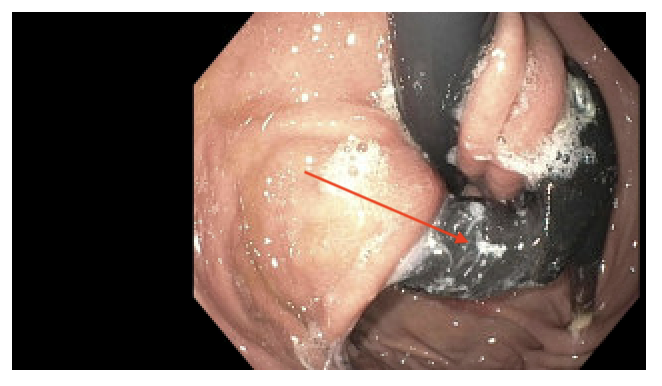
Figure: Endoscopy showing gastric band eroded into stomach (Red arrow).
Disclosures: Karan Yagnik indicated no relevant financial relationships. Suryansh Atreya indicated no relevant financial relationships. Pranay Joshi indicated no relevant financial relationships. Sankalp Acharya indicated no relevant financial relationships. Dhramesh Kaswala indicated no relevant financial relationships. Wael Ghali indicated no relevant financial relationships.
Karan J.. Yagnik, MD, Suryansh Atreya, MD, Pranay Joshi, MD, Sankalp Acharya, MD, Dhramesh Kaswala, MD, Wael Ghali, MD. P2137 - Twenty Years Later, Trouble Returns: Late-Onset Gastric Band Erosion Unmasked, ACG 2025 Annual Scientific Meeting Abstracts. Phoenix, AZ: American College of Gastroenterology.

 photo")